

What Is Schwannoma (Nerve-Sheath Tumor)? Causes, Symptoms, and Treatment


What Is a Schwannoma (Nerve-Sheath Tumor)?
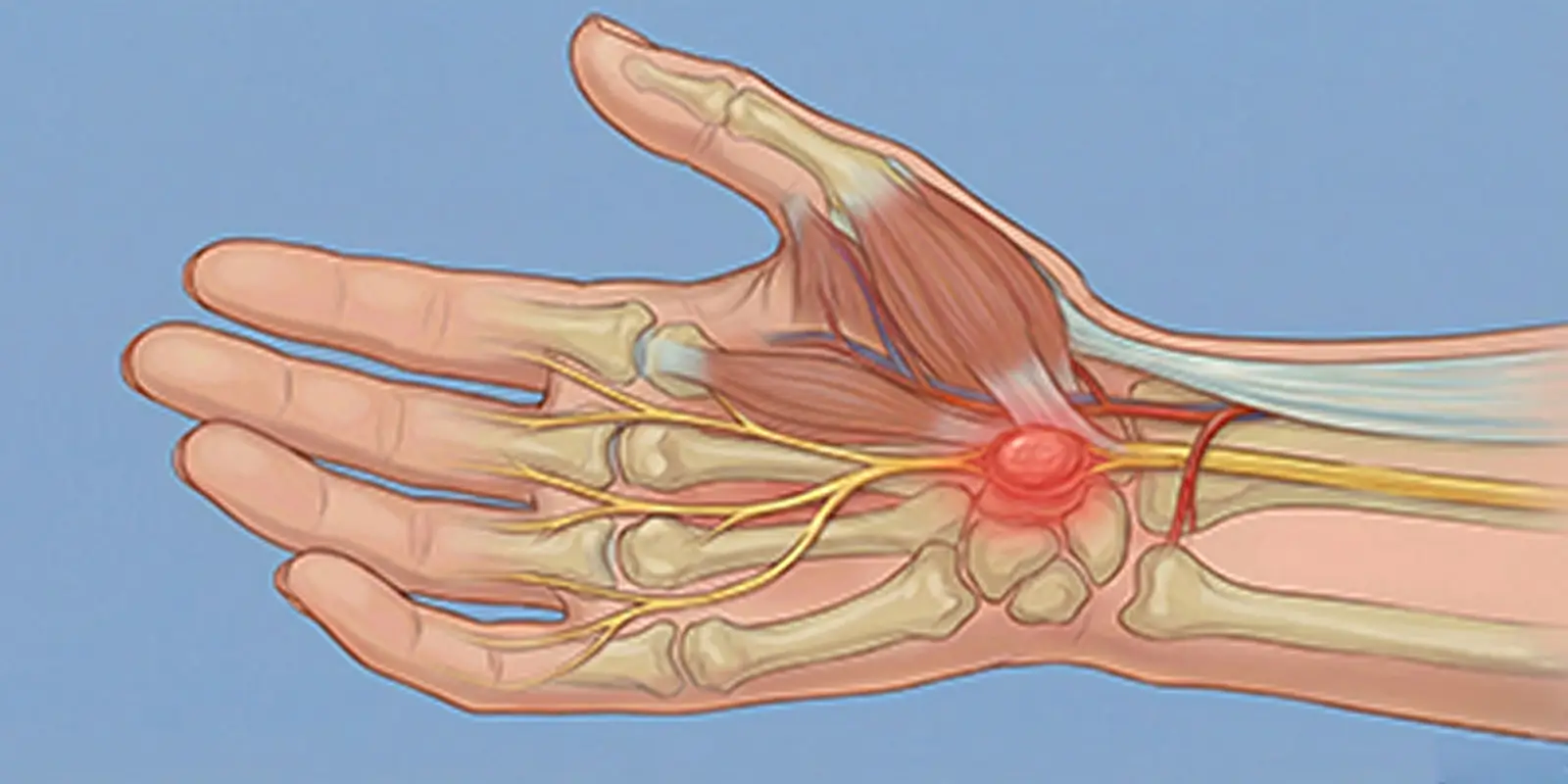
A schwannoma is a nerve-sheath tumor that arises from Schwann cells, which wrap nerves and enable conduction, and that usually follows a benign course. These tumors can develop on peripheral nerves, spinal nerve roots, and cranial nerves. Because they tend to grow slowly, they may cause no symptoms for a long time; as they enlarge, however, they can press on the nerve and cause serious neurologic problems.
In orthopedic oncology practice, schwannomas can be confused with a bone tumor and with soft-tissue tumors, especially when they sit in the spine and deep tissues. Correct diagnosis is therefore critical for avoiding unnecessary surgery and protecting nerve tissue.
According to the clinical approach emphasized by Prof. Dr. Yavuz Arıkan, detecting schwannomas early allows effective treatment to be planned before nerve injury occurs.
What Kind of Tumor Is a Schwannoma?
Schwannomas are usually encapsulated, arise from a single nerve, grow slowly, and are mostly benign. Because of these features they do not spread into nearby tissues, but they can compress the nerve and cause pain and loss of function.
Difference Between Schwannomas and Malignant Tumors
Schwannomas are usually not cancer. In very rare cases they can be confused with malignant peripheral nerve-sheath tumors. This distinction is made especially with MRI and, when needed, biopsy. From an orthopedic oncology standpoint this distinction is vital.
Why Does It Matter?
When nerve tissue is damaged, lasting numbness, muscle weakness, and loss of movement can develop. Although a schwannoma is benign, it is therefore a nerve-sheath tumor that should not be neglected.
Why Does a Schwannoma Form?
A schwannoma (nerve-sheath tumor) appears when Schwann cells that wrap nerves multiply in an uncontrolled way. These cells normally insulate nerve fibers and regulate electrical conduction. In some situations cellular control mechanisms fail, and these cells multiply abnormally and form a tumor. Clinical observations in orthopedic oncology show that schwannoma development depends not on a single cause but on several biologic factors.
1. Genetic Mutations
The most basic cause of schwannomas is genetic changes in Schwann cells. Disruptions in genes that control cell growth can cause these cells to multiply without limit.
2. Neurofibromatosis Type 2 (NF2)
Some schwannomas are related to a hereditary disease called neurofibromatosis type 2. In this disease, mechanisms that suppress tumor formation in nerve-sheath cells do not work, and more than one schwannoma can develop.
3. Radiation Exposure
In areas that previously received radiotherapy, the risk of a schwannoma developing years later can increase. Radiation can damage cell DNA and trigger tumor formation.
4. Nerve Trauma and Chronic Irritation
In some cases, schwannoma development is seen more often in nerves that were previously injured or are under constant pressure. Long-term mechanical stress can disrupt cellular repair processes.
5. Sporadic (Incidental) Development
A large proportion of schwannomas appear completely by chance, without any genetic disease. In these patients no specific triggering factor can be found.
Where Do Schwannomas Occur?
Because a schwannoma (nerve-sheath tumor) develops from Schwann cells that wrap nerves, it can appear almost anywhere in the body. In orthopedic oncology practice, however, it is most common in the spine, arm and leg nerves, and the head and neck. In some sites these tumors are noticed only as a small swelling; in others they can cause serious pain and loss of movement.
In clinical evaluations by Prof. Dr. Yavuz Arıkan, schwannomas can, depending on location, be confused with a bone tumor or soft-tissue masses, and diagnosis of schwannomas around the spine in particular requires great care.
1. Spine and Spinal Nerve Roots
The most common site of schwannomas is the spine. Schwannomas that develop from nerve roots in the neck, back, and lower back can present with neck and back pain, pain radiating to the arm or leg, numbness, and muscle weakness. These locations can sometimes raise suspicion of a bone tumor on imaging.
2. Head and Facial Nerves
Schwannomas that arise especially from the hearing nerve (vestibular schwannoma) cause symptoms such as hearing loss, tinnitus, and balance problems. Tumors near the facial nerve can lead to weakness of the facial muscles.
3. Arm and Hand
Schwannomas that develop in nerves around the elbow, forearm, and wrist present as a soft mass that can be felt, pain that increases with touch, and numbness in the fingers. These masses can sometimes be thought to originate from soft tissue or bone.
4. Leg and Foot Nerves
Schwannomas around the thigh, behind the knee, and the ankle can cause pain while walking, an electric-shock-like sensation, and weakness.
5. Abdomen and Pelvis
Schwannomas that arise from deep nerves can develop inside the abdomen or pelvis. These tumors can present with pressure on organs and unexplained abdominal or groin pain, and on imaging they can be confused with a bone tumor or other masses.
What Are the Symptoms of a Schwannoma?
Because a schwannoma develops from the nerve sheath, symptoms depend directly on the function of the affected nerve. Some patients therefore have only a mild feeling of discomfort, while others have serious pain and loss of function. Schwannomas on the spine and large nerves in particular can over time disrupt nerve conduction and clearly affect daily life.
1. Pain Spreading Along the Nerve
The most common symptom of a schwannoma is pain felt along the path of the nerve. This pain can radiate from the neck to the arm, from the lower back to the leg, or from the shoulder to the hand. Patients often describe this pain as burning or an electric shock.
2. Numbness and Tingling
As the tumor presses on the nerve, numbness, tingling, and reduced sensation develop in the areas that nerve supplies. These signs usually progress slowly and may not be noticed at first.
3. Muscle Weakness
In schwannomas that affect motor nerves, weakness in the arm or leg, difficulty holding objects, and impaired walking balance can appear. This is especially noticeable in spine-origin schwannomas.
4. A Palpable Mass
Schwannomas that develop from superficial nerves in the arm, leg, or neck can be felt as a slowly growing, mobile mass under the skin that is tender when pressed. These masses can sometimes be confused with a formation of bone or soft-tissue origin.
5. Night Pain and Discomfort at Rest
In some schwannomas, pain becomes more noticeable especially at night or at rest. This can disrupt sleep quality.
6. Findings Specific to Spinal Schwannomas
Schwannomas near the spinal cord and nerve roots can cause more serious neurologic symptoms such as lower-back and neck pain, walking difficulty, and changes in bladder or bowel control.
How Is a Schwannoma Diagnosed?
Diagnosis of a schwannoma (nerve-sheath tumor) is not made simply by seeing a mass; it is made by correctly showing whether that mass truly originates from a nerve. Depending on location, schwannomas can be confused with a bone tumor, a disc herniation, an intramuscular mass, or other soft-tissue tumors. The diagnostic process therefore includes both clinical evaluation and advanced imaging.
1. Detailed Clinical Evaluation
The first step is analysis of the patient’s complaints. Pain spreading along a nerve path, numbness and tingling, muscle weakness, and a tender palpable mass are warning findings for a schwannoma. The physician determines by examination which nerve these symptoms belong to.
2. Magnetic Resonance Imaging (MRI)
MRI is the most valuable method for diagnosing a schwannoma. MRI clearly shows whether the tumor originates from a nerve, its relationship to the nerve, and its connection to nearby muscle, bone, and vessels. The encapsulated structure of schwannomas and their continuity with the nerve are distinctive features on MRI.
3. Computed Tomography (CT)
CT is used especially for schwannomas next to the spine and bone. Whether the tumor is pressing on bone and whether it has widened bone canals is assessed. This allows distinction from suspicion of a bone tumor.
4. Ultrasound
Ultrasound is very useful for schwannomas in superficial areas such as the arm, leg, and neck. A well-defined mass related to a nerve suggests a schwannoma.
5. Electrophysiologic Tests
In some patients, EMG (electromyography) is used to measure nerve conduction. This test shows whether there is pressure or damage in the nerve.
6. Biopsy (When Needed)
Most schwannomas do not need a biopsy. If imaging cannot exclude a malignant tumor, or if the mass is growing rapidly, a controlled biopsy is done. Biopsy of nerve-origin tumors requires a special technique.
Treatment Methods
Schwannoma (nerve-sheath tumor) treatment is planned according to the nerve involved, the growth rate, the complaints it causes, and its relationship to nearby tissues. The aim is not only to eliminate the tumor but also to preserve nerve function. Schwannoma treatment therefore requires orthopedic oncology and nerve surgery to be considered together, unlike an ordinary mass operation.
1. Observation and Active Surveillance
For small, slow-growing, asymptomatic schwannomas, the first option is often observation without treatment. During this process, regular MRI, neurologic examination, and monitoring of pain and sensory changes are done. If the tumor does not grow or press on the nerve, surgery may not be needed.
2. Medication and Symptom Control
In patients who have pain but do not yet need surgery, drugs that reduce nerve pain and treatments that suppress inflammation can be used. This approach helps protect quality of life, especially in spinal schwannomas.
3. Surgery
Surgery is the most effective option in the following situations: severe or progressive pain, numbness and muscle weakness, growth of the tumor, and deterioration of nerve function.
During surgery the aim is to separate the schwannoma carefully from the nerve and remove it. Because schwannomas are usually encapsulated, they can be removed while preserving the nerve in experienced hands. This feature distinguishes them from many bone tumors or aggressive soft-tissue tumors.
4. Radiosurgery and Focused Radiotherapy
In some patients, if surgical risk is high or the tumor sits in a difficult location, radiosurgery may be preferred. Especially in vestibular schwannomas, this method can control growth.
5. Rehabilitation After Surgery
Because nerve tissue is sensitive, physical therapy after surgery, monitoring of nerve function, and muscle-strength exercises are very important. This process plays a critical role in preventing lasting weakness.
Difference Between a Schwannoma and a Bone Tumor
A schwannoma (nerve-sheath tumor) and a bone tumor can resemble each other in both imaging findings and the patient’s complaints, especially when they appear in the spine and deep tissues. The two diseases, however, are completely different in origin, behavior, and treatment. Understanding this difference correctly is very important for preventing misdiagnosis and unnecessarily aggressive treatment.
1. Tissue of Origin
A schwannoma develops from Schwann cells that wrap nerves. It is therefore a nerve-origin tumor. A bone tumor arises directly from bone tissue, bone marrow, or cartilage cells. This basic difference determines how the tumor behaves in the body.
2. Growth Pattern
Schwannomas usually grow slowly, are encapsulated, and grow by pushing the nerve from the outside. Bone tumors develop from inside the bone, progress by destroying or hardening bone tissue, and can weaken nearby bone.
3. Pain and Clinical Findings
In schwannomas, pain usually spreads along the nerve, is like an electric shock or burning, and can change with position and movement. In bone tumors the pain is deeper and duller, tends to increase at night, and may be accompanied by swelling and tenderness.
4. Imaging Differences
On MRI and CT, schwannomas appear as well-defined, encapsulated masses along the nerve path. Bone tumors appear as lesions that disrupt, expand from within, or destroy bone structure. Schwannomas around the spine can press on bone and look like a bone-origin problem, which leads to confusion.
5. Risk of Being Malignant
The large majority of schwannomas are benign. Bone tumors may be benign or malignant, and some types can threaten life.
6. Treatment Approach
In schwannoma treatment the priority is to remove the tumor while protecting the nerve. In bone tumors, wide surgery, bone reconstruction, and sometimes chemotherapy or radiotherapy may be needed. Mistaking a schwannoma for a bone tumor can therefore put the patient through an unnecessarily heavy treatment process.
Conclusion
A schwannoma (nerve-sheath tumor) develops from cells that wrap nerves and often progresses slowly, but depending on location it can seriously affect quality of life. Schwannomas that develop in the spine, arm and leg nerves, the head and neck, or nerves inside the abdomen can present with very different symptoms such as pain, numbness, muscle weakness, and limited motion. This wide range of symptoms can cause the disease to be confused sometimes with a simple musculoskeletal problem and sometimes with a bone tumor.
In diagnosis, detailed clinical evaluation, MRI, and CT when needed are used together to clarify whether the tumor truly originates from a nerve or from bone or soft tissue. This distinction is critical both for preventing unnecessary heavy treatments and for protecting nerve tissue. The treatment plan is determined according to the tumor’s size, growth rate, and the effects it causes; some patients are only observed, while others need surgery or focused radiotherapy.
In the orthopedic oncology approach adopted by Prof. Dr. Yavuz Arıkan, the aim is not only to eliminate the schwannoma but to protect the patient’s nerve function and daily quality of life. With correct diagnosis, appropriate treatment at the right time, and regular follow-up, the large majority of patients with a schwannoma can reach safe, successful long-term results.
Frequently Asked Questions
Frequently Asked Questions
A schwannoma is a usually benign tumor that develops from Schwann cells that wrap nerves. Because it grows along the nerve, it can cause pain, numbness, and weakness, and it can sometimes be confused with a bone tumor.
The large majority are benign. In very rare cases it can transform into a malignant nerve-sheath tumor. Regular follow-up and imaging are therefore important.
They most often appear around the spine, in arm and leg nerves, in the head and neck, and in nerves inside the pelvis. In these sites they can press on bone and mimic bone-origin pain.
Yes. Pain usually spreads along the nerve path and is felt as burning, stabbing, or an electric shock. It can increase or decrease with movement.
No. A schwannoma is nerve in origin; a bone tumor arises from bone tissue. Around the spine and in deep tissues, however, the two can look similar on imaging.
Magnetic resonance imaging (MRI) is the most important imaging method. CT and, rarely, biopsy are used when needed for a definitive diagnosis. An orthopedic oncology approach is very important in diagnosis.
No. Small, asymptomatic, non-growing schwannomas can be followed with regular observation. Those that cause pain, neurologic loss, or rapid growth require surgery.
Because it is related to a nerve, it requires experience. With appropriate technique, nerve function is preserved in most patients. It should therefore be done at specialized centers.
The chance of recurrence of a completely removed schwannoma is low. Rarely, however, it can grow again, so follow-up is needed.
No. Because most schwannomas are benign, with correct diagnosis and appropriate treatment patients can have a normal lifespan and quality of life. In the multidisciplinary approach used by Prof. Dr. Yavuz Arıkan, both tumor control and nerve health are protected together.
 WhatsApp
  Facebook
  Twitter
  Linkedin



 +90 505 398 34 34
 doctoryavuzarikan@gmail.com
 Book Appointment / Contact