

What Is a Comminuted Fracture?


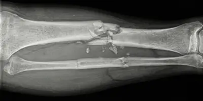
What Is a Comminuted Fracture?
A comminuted fracture is one of the most serious fracture types, occurring when a bone is divided into more than two pieces. In medical language this is called a “comminuted fracture,” meaning that the integrity of the bone is completely disrupted and the fragments are separated from each other. Unlike simple fractures, the bone is broken into multiple small pieces, so it is extremely difficult for it to heal correctly on its own.
Comminuted fractures usually result from high-energy trauma. Traffic accidents, falls from height, workplace accidents, and severe sports injuries are the most common causes. In people with diseases that reduce bone density, such as osteoporosis, even lower-intensity impacts can lead to a comminuted fracture. In older people these fractures can therefore be more common and more severe.
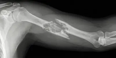
Comminuted fractures are most often seen in the thigh bone (femur), upper arm bone (humerus), shin bone (tibia), and around the wrist. These regions bear load and are exposed to impact, so they are at high risk. When the fracture occurs, the bone ends displace and surrounding muscles, vessels, and nerves can be injured. This makes comminuted fractures not only a bone injury but also a serious soft-tissue trauma.
The most important feature of comminuted fractures is that they cannot heal with a simple cast or rest. Surgery is usually needed to bring the fragments into the correct position and fix them. Otherwise the bone may heal incorrectly, and deformity, limited motion, and permanent disability can develop.
In summary, a comminuted fracture is an orthopedic trauma that requires emergency care and specialist skill. With early diagnosis, correct surgery, and good rehabilitation, patients can largely return to their previous lives. Delayed or inadequate treatment, however, can lead to permanent loss of function.
Why Does a Comminuted Fracture Occur?
A comminuted fracture occurs when the force applied to the bone exceeds its resistance. These fractures usually form with high-energy trauma and cause the bone to break into more than two pieces. Both external factors and weakness of bone structure play an important role.
One of the most common causes is traffic accidents. High-speed trauma such as motorcycle accidents, in-vehicle collisions, and pedestrian impacts places a large force on the bone and leads to a comminuted fracture. In these accidents not only bone but also surrounding muscle, vessels, and nerves can be injured.
Falls from height are also among the most important causes. In falls from stairs, a balcony, or a construction site, the body’s entire weight can land on a single bone and the bone can break into many pieces. The feet, legs, and hip bones are often affected in these accidents.
Workplace accidents and severe trauma also increase comminuted fracture risk. Crushing, compression, and falls in construction, industry, and jobs that involve carrying heavy loads can fragment the bone. In athletes, high-contact sports, sudden collisions, and uncontrolled falls can result in a comminuted fracture.
In addition, diseases that weaken bone tissue, such as osteoporosis (bone thinning), make a comminuted fracture more likely. In people with reduced bone density, a simple fall or bump can cause the bone to break into more than two pieces. In older people these fractures can therefore be more common and more severe.
In some cases, diseases such as bone tumors or bone metastases also weaken bone and increase comminuted fracture risk. These fractures are called pathologic fractures, and treatment of the underlying disease is also very important.
What Are the Symptoms of a Comminuted Fracture?
Symptoms of a comminuted fracture are much more severe and obvious than those of simple fractures. Because the bone is broken into more than two pieces, surrounding muscle, vessel, and nerve tissues can be seriously injured. Comminuted fractures are therefore among orthopedic traumas that require emergency care.
The most obvious symptom is severe, persistent pain. This pain usually increases with movement and does not fully go away even with rest. Intense tenderness develops in the fracture region and the patient cannot use that limb. In comminuted fractures of the leg or arm especially, the patient cannot bear weight or lift the arm.
Swelling and bruising are other important symptoms. After the fracture, bleeding and swelling form in the region, quickly leading to obvious swelling and bruises under the skin. As swelling increases, the skin tightens and pain becomes even more severe.
Deformity and abnormal posture are among the most striking findings. Because the fragments have displaced, the arm, leg, or affected limb may lose its normal shape and look bent or twisted. In some cases the bone ends can be felt under the skin, or if there is an open fracture they can be seen from outside.
Loss of motion is also an important symptom. Movement in the comminuted fracture region becomes almost impossible. The patient cannot even do simple movements and wants to keep the limb still because of severe pain.
In severe comminuted fractures, circulation problems such as numbness, coldness, or color change may also be seen. This can show that vessels or nerves were injured during the fracture and requires emergency care.
In summary, comminuted fracture symptoms present with severe pain, swelling, deformity, loss of motion, and circulation problems. If even one of these symptoms is present, going to the emergency department without delay and being evaluated by an orthopedic specialist is vital.
How Is a Comminuted Fracture Diagnosed?
Correct, rapid diagnosis of a comminuted fracture is very important both for reducing the risk of permanent disability and for determining the most appropriate treatment. Because the bone is broken into more than two pieces, a wrong or incomplete diagnosis can lead to serious complications.
The diagnostic process starts first with the history of the trauma. A history of a traffic accident, a fall from height, or a severe impact strongly suggests a comminuted fracture. The physician then assesses pain, swelling, deformity, and loss of motion in the fracture region with physical examination. Examination alone, however, cannot clearly determine the type of fracture.
The most basic imaging method is X-ray. X-ray clearly shows the number of fragments, the degree of displacement, and which bone is affected. In comminuted fractures, X-ray clearly shows that the bone has separated into multiple fragments.
For more detailed assessment, computed tomography (CT) is often obtained. CT shows the three-dimensional structure of the fracture, revealing small fragments, fractures that extend to the joint surface, and the need for surgery much more clearly. These images are vital especially for surgical planning.
In some cases magnetic resonance imaging (MRI) may also be requested. MRI shows how much damage there is to soft tissues such as muscles, ligaments, tendons, and nerves together with the bone. In comminuted fractures around the knee, shoulder, or wrist especially, MRI helps detect additional injuries.
In severe comminuted fractures, if vessel or nerve injury is suspected, additional tests such as vascular ultrasound or angiography may also be done. Circulation of the limb is thereby assessed and vital risks are recognized early.
In summary, diagnosis of a comminuted fracture is made by using advanced imaging methods such as X-ray, CT, and, when needed, MRI together. Correct diagnosis is the basis of correct surgery and a successful recovery.
How Is a Comminuted Fracture Treated?
Treatment of a comminuted fracture is completely different from that of simple fractures and must be planned by a specialist in orthopedics and traumatology. Because the bone is broken into more than two pieces, it cannot heal correctly on its own. The main aim of treatment is to bring the fragments into the correct position, fix the bone solidly, and enable the patient to move safely as soon as possible.
In the first stage, the patient’s general condition is assessed. In severe trauma such as a traffic accident or a fall from height, internal organ injuries may also be present, so vital risks are controlled first. The fracture region is temporarily immobilized with a splint or fixer and pain is controlled.
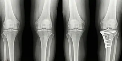
The great majority of comminuted fractures require surgical treatment. The reason is that the fragments cannot be held in the correct position with a cast. During surgery the fragments are placed one by one in their anatomic position and fixed with plate, screw, nail, or wire systems. This method allows the bone to heal correctly and eliminates the risk of malunion.
In some cases, if there is bone loss or serious fragmentation, a bone graft (support taken from the patient’s own bone or from artificial bone tissue) is used. This helps the bone heal more strongly.
After surgery, a controlled weight-bearing and early movement program is usually applied. In this process, physical therapy and rehabilitation are very important for strengthening muscles, preserving joint motion, and helping the patient return to daily life in a healthy way.
Regular X-ray checks are done throughout treatment. These checks show whether the bone is healing correctly. If healing is insufficient, additional interventions may be needed.
How Is Comminuted Fracture Surgery Performed?
Comminuted fracture surgery is a surgical procedure used in serious fractures in which the bone is broken into more than two pieces, aiming to make the bone solid and functional again. The main goal is to bring the fragments into the anatomically correct position, fix them stably, and allow the fracture to heal healthily.
Before surgery, detailed imaging is done. With X-ray and especially computed tomography (CT), how many pieces the bone has broken into, whether the fracture extends to the joint surface, and how much the bone is damaged are assessed. Based on these images, the type of surgery and the size and placement of the plates, screws, or nails to be used are planned.
Surgery is usually done under general anesthesia. The surgeon reaches the fracture region and seats the fragments one by one. Correct alignment of the fragments at this stage is extremely critical, because even the smallest error can lead to deformity or limited motion in the bone.
After the fragments are brought into the correct position, the bone is fixed solidly using plates and screws, intramedullary nails, or special fixer systems.
If there is serious fragmentation or bone loss, the gaps are filled with a bone graft or synthetic bone filler. This helps the bone heal more strongly and quickly.
After surgery the fracture region is usually supported with a cast or splint. Thanks to modern surgical techniques, however, early movement becomes possible in most patients. This prevents muscle loss and speeds recovery.
One of the most important stages after comminuted fracture surgery is also physical therapy and rehabilitation. With correct exercises, joint motions are preserved, muscles are strengthened, and the patient can return safely to daily life.
Recovery After a Comminuted Fracture
Recovery after a comminuted fracture is a period that needs to be managed longer and more carefully than after simple fractures. Because the bone is broken into more than two pieces, healing of both the bone and surrounding soft tissues takes time. Success of this process is directly related to the quality of the surgery, the patient’s general health, and adherence to rehabilitation.
In the first days after surgery the aims are to control pain, reduce swelling, and protect the fracture region. A splint or supportive fixer is usually applied. Starting controlled movements early as recommended by the physician is very important for preventing joint stiffness and muscle wasting.
Within the first 4–6 weeks, the bone healing process begins. In this period the patient moves with crutches or support devices without putting full weight on the fracture region. Regular X-ray checks follow whether bone healing is progressing healthily.
Physical therapy and rehabilitation are the most critical stage of recovery. Individualized exercise programs are applied to regain muscle strength, preserve range of motion, and restore normal walking or arm use. The more regularly and disciplined this period is, the lower the risk of permanent limited motion.
Bone healing is usually completed between 3 and 6 months. Return to sport, carrying heavy loads, or intensive physical activity may take longer. In some patients full recovery may take 9–12 months.
During recovery, smoking, inadequate nutrition, and inactivity negatively affect bone healing. A diet rich in protein, calcium, and vitamin D and adherence to the lifestyle changes recommended by the physician are therefore very important.
Complications of Comminuted Fractures
Because comminuted fractures are traumas in which bone integrity is seriously disrupted, they carry a much higher complication risk than simple fractures. If not treated correctly and in time, these complications can lead to permanent loss of motion, deformity, and chronic pain.
One of the most common complications is nonunion or delayed union. If the fragments are not fixed well enough or blood supply is disrupted, the bone does not heal in the desired time. This can cause ongoing pain and loss of function and may require additional surgery.
Malunion is also an important problem. If the fragments heal without correct alignment, the bone may heal bent or short. This can lead to gait disturbance, joint problems, and aesthetic deformities.
In comminuted fractures, surrounding vessels and nerves can be injured. These injuries may present with numbness, weakness, coldness, or circulation problems. If untreated, permanent damage can occur.
Infection is an important risk especially in open comminuted fractures and after surgery. Infected bone tissue does not heal and may require long-term antibiotics or another operation.
In comminuted fractures near a joint, joint stiffness and osteoarthritis (arthrosis) can develop. This causes limited motion and chronic pain in the long term.
Systemic problems related to prolonged inactivity, such as muscle wasting, clot formation, and joint stiffness, may also be seen.
What Should Patients With a Comminuted Fracture Watch For?
For patients who have had a comminuted fracture, treatment does not end with surgery. Real success is determined by how well the patient adheres to the process after surgery. Measures taken in daily life and adherence to the physician’s recommendations are therefore very important for healthy bone healing.
First, the patient must strictly follow the weight-bearing instructions given by the physician. Putting early, excessive load on the fractured leg or arm can displace plates and screws and disrupt bone healing. Supportive devices such as crutches, a walker, or a sling must be used for the recommended period.
Staying away from smoking and tobacco products is very important. Nicotine reduces blood supply to bone tissue and seriously delays fracture healing. In patients who smoke, nonunion risk is clearly higher.
Nutrition also plays a critical role. A diet rich in protein, calcium, vitamin D, and zinc speeds repair of bone tissue. Dairy products, green leafy vegetables, eggs, fish, and legumes should definitely be part of the diet in this process.
Physical therapy and exercise programs should not be skipped. Remaining inactive leads to muscle wasting, joint stiffness, and long-term loss of motion. Exercises done under specialist supervision both increase muscle strength and preserve normal range of motion of the joints.
It is also very important to attend regular physician visits and follow bone healing with X-rays. If unusual symptoms such as pain, swelling, redness, or sudden loss of function appear, a physician should be contacted without delay.
Does a Comminuted Fracture Leave Permanent Damage?
Comminuted fractures are severe traumas that can cause serious damage to bone and surrounding tissues. Many patients therefore wonder whether permanent damage will occur after this type of fracture. The risk of permanent damage depends on the severity of the fracture, its location, the quality of the treatment applied, and adherence during recovery.
With timely surgical intervention, anatomic placement of the fragments, and use of strong fixation systems, the risk of permanent disability can be substantially reduced. Thanks to modern orthopedic surgical techniques, a large part of patients can return to their previous mobility and daily life functions.
In some cases, however, permanent damage can develop. Especially in comminuted fractures that extend to the joint surface, if joint cartilage has been damaged, osteoarthritis (arthrosis) may appear in later years. This can cause stiffness, pain, and limited motion in the joint.
In severe comminuted fractures accompanied by vessel or nerve injury, permanent numbness, loss of strength, or circulation problems may be seen in the limb. Shape problems such as malunion, curvature, or shortening of the bone can also lead to gait disturbance and posture problems in the long term.
The most important way to reduce the risk of permanent damage is early diagnosis, correct surgery by an experienced surgeon, and a disciplined rehabilitation process. Long-term results are much more successful in patients who continue physical therapy regularly and follow the physician’s weight-bearing and movement recommendations.
Conclusion
A comminuted fracture is one of the most severe fracture types, in which the bone is broken into more than two pieces by high-energy trauma. Traffic accidents, falls from height, and workplace and sports accidents are among the most common causes. In people with osteoporosis or bone diseases, even smaller impacts can lead to a comminuted fracture. Comminuted fractures are therefore not only a bone injury but also a serious trauma that can affect the whole body.
Comminuted fractures that present with severe pain, obvious swelling, deformity, and loss of motion must be evaluated with advanced imaging. The structure of the fracture is analyzed in detail with X-ray and computed tomography, and an individualized plan is created. The great majority of comminuted fractures require surgical treatment, because the bone must be reconstructed with fixer systems such as plates, screws, or nails so that the fragments heal correctly.
The recovery process after surgery is as important as the surgery itself. Regular checks, correct weight-bearing, physical therapy, and healthy lifestyle habits allow the bone to heal solidly. Otherwise, serious complications such as nonunion, malunion, infection, and joint stiffness can develop. Adherence to rehabilitation especially directly affects the patient’s long-term function.
Although comminuted fractures have the potential to leave permanent damage, thanks to modern surgical techniques and a disciplined treatment process, a large part of patients can return to normal life. Early diagnosis, correct surgery by an experienced orthopedic team, and effective rehabilitation substantially reduce the risk of permanent disability.
Frequently Asked Questions
Frequently Asked Questions
A comminuted fracture is a serious fracture in which the bone is broken into three or more pieces, not two. It usually occurs after traffic accidents, falls from height, and severe trauma.
No. Comminuted fractures usually require surgery. The bone fragments must be fixed with plates, screws, or a nail so that they heal in the correct position.
In most cases yes. Because bone stability is lost in comminuted fractures, surgery is usually unavoidable. Surgery provides correct alignment and solid healing of the bone.
Healing time is usually between 3 and 6 months. The location of the fracture, the patient’s age, and general health can affect this period.
Yes. With correct surgery, regular physical therapy, and rehabilitation, most patients can walk normally again.
If timely, correct treatment is not applied, there is a risk of permanent damage. With modern orthopedic techniques, this risk can be substantially reduced.
Pain is normal in the first weeks. As painkillers are used and treatment progresses, pain gradually decreases.
They can often stay. If they cause discomfort or a complication develops, they can be removed in later years.
Absolutely yes. Physical therapy is very important for regaining muscle strength and preventing joint stiffness.
After the bone has fully healed, the risk of fracturing again is similar to that of normal bones. If the bone remains weak, however, the risk may increase.
 WhatsApp
  Facebook
  Twitter
  Linkedin



 +90 505 398 34 34
 doctoryavuzarikan@gmail.com
 Book Appointment / Contact