

What Is Osteochondroma (Bone Cancer)? Symptoms, Risks, and Treatment


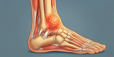
Osteochondroma is the most common benign bone tumor, developing from bone and cartilage. It usually appears in childhood and adolescence and grows near the ends of long bones. Although it is considered benign, some cases can transform into a malignant bone cancer called chondrosarcoma, so careful follow-up is required.
Prof. Dr. Yavuz Arıkan, who has worked in orthopedic oncology for many years, emphasizes that osteochondromas that are not followed correctly can progress to a sarcoma picture, and that early diagnosis is therefore vital.
What Is Osteochondroma?
Osteochondroma is a growth that develops from the outer surface of bone, is covered by a cartilage cap, and is considered the most common benign bone tumor. It usually appears in childhood and adolescence and is seen near the growth plates of long bones.
The most common sites are:
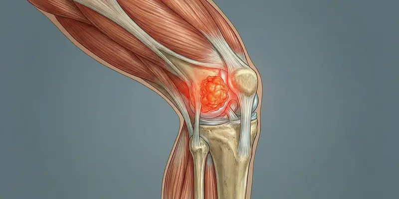
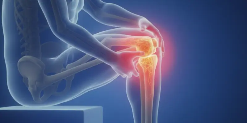
- Around the knee (femur – thigh bone and tibia – shinbone)
- Shoulder and upper arm bone (humerus)
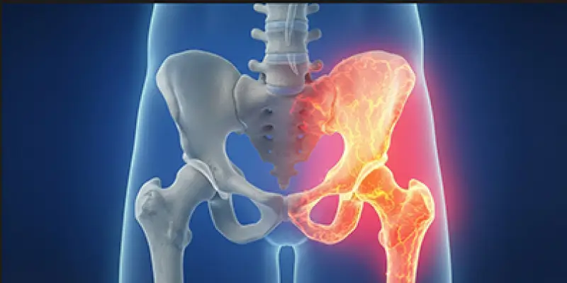
- Hip and pelvis
- Leg bones
Osteochondromas often cause no pain and are noticed only as a hard mass. As they grow, they can press on nearby muscles, tendons, nerves, and vessels and cause symptoms. Osteochondromas near a joint can restrict motion.
According to Prof. Dr. Yavuz Arıkan, even though osteochondromas are considered benign, their biologic behavior should be watched carefully. Some osteochondromas can change cellular structure over time and become more aggressive tumors.
Is Osteochondroma Bone Cancer?
Osteochondroma is not bone cancer at the outset. Medically it is classified as a “benign bone tumor.” In a defined group of patients and under certain conditions, however, osteochondromas can transform into a malignant bone tumor called chondrosarcoma. Chondrosarcoma is a type of sarcoma, that is, a bone cancer.
This transformation is seen especially when:
- The osteochondroma continues to grow in adulthood
- Pain increases at night and does not ease with rest
- The mass hardens and changes shape
- MRI shows thickening of the cartilage cap
- Size increases rapidly
These findings indicate that the osteochondroma may no longer be only a benign prominence and may be entering a process of malignant change.
Such suspicious osteochondromas are evaluated with advanced imaging and biopsy. Prof. Dr. Yavuz Arıkan emphasizes that adult osteochondroma patients are at higher risk in this regard and must be followed by a specialist orthopedic oncologist.
Osteochondroma Symptoms
Most osteochondromas cause no symptoms at first. As they grow, the following complaints can appear:
- A hard, fixed mass on the bone
- Regional pain that increases with movement or is constant
- Limited joint motion
- Numbness or tingling from nerve compression
- Visible deformity
If pain increases at night, the mass grows rapidly, or sudden tenderness develops, this may be an early sign of sarcoma transformation.
Why Does Osteochondroma Form?
The cause cannot be tied to a single factor in every patient. In most cases the tumor appears when cartilage cells deviate from the normal growth plate during bone growth and multiply in an uncontrolled way toward the outside of the bone. Osteochondromas therefore develop most often in childhood and adolescence, when bones lengthen rapidly.
Normally, cartilage cells in the growth plate allow the bone to lengthen. In some people these cells multiply in the wrong direction and form a prominence extending from the bone surface. This structure later ossifies and becomes an osteochondroma.
Genetic Factors and Hereditary Predisposition
Some osteochondromas have a genetic origin. In multiple hereditary osteochondromatosis (MHE), also called hereditary multiple exostoses, many osteochondromas develop in various parts of the body. This disease is autosomal dominant; if one parent is a carrier, the chance of it appearing in a child is high.
In patients with MHE:
- The number of osteochondromas is high
- Tumors appear at a younger age
- Deformity and limb shortening are more common
- The risk of chondrosarcoma (bone cancer) is much higher than in people without MHE
The rate of malignant transformation of osteochondromas in these patients is reported as 5–25%. This is considerably higher than in people with a single osteochondroma.
Why Do Some Transform Into Cancer?
Osteochondroma cells can accumulate genetic mutations over time. In patients with MHE, disorders in the EXT1 and EXT2 genes lead to uncontrolled cell growth. These cellular changes set the stage for the osteochondroma to transform into a sarcoma type such as chondrosarcoma.
According to Prof. Dr. Yavuz Arıkan, osteochondromas that continue to grow in adulthood or cause pain may already be losing their benign character at the cellular level.
Trauma and Environmental Factors
Although osteochondromas mainly originate from the growth plate, in some cases osteochondroma-like prominences can develop after:
- Bone trauma in childhood
- Disorders of bone development
- Previous surgery
These are usually different from true osteochondromas and have a lower risk of malignant transformation.
How Is Osteochondroma Diagnosed?
Diagnosis is not made simply by seeing a mass. The aim is not only to answer “Is this an osteochondroma?” but also to clarify whether there is a risk of malignant transformation. The diagnostic process should therefore follow orthopedic oncology principles.
1. Clinical Examination
Patients usually present after noticing a hard, immobile mass on the bone. Examination assesses:
- Firmness of the mass
- Tenderness to pain
- Relationship to movement
- Signs of nerve or vessel compression
Night pain, rapid growth, or new tenderness is a warning for sarcoma.
2. X-ray
This is the first and most important step in diagnosis. On X-ray:
- A prominence extending outward from the bone
- A structure connected to the parent bone
- Typical bone density
is seen. This appearance is often diagnostic for osteochondroma.
3. Magnetic Resonance Imaging (MRI)
MRI shows the cartilage cap, the most critical component of osteochondroma. The thickness of this cap is the most important indicator of malignant transformation.
- Less than 1 cm → generally safe
- Greater than 2 cm → chondrosarcoma risk increases
MRI also assesses:
- Soft-tissue extension
- Relationship to vessels and muscle
- Edema and invasion
4. Computed Tomography (CT)
CT shows bone structure in detail. It is preferred especially for osteochondromas of the pelvis, hip, and spine.
5. Biopsy (If Needed)
If imaging is suspicious, biopsy is required. If biopsy is done with the wrong technique, cancer cells can spread.
Prof. Dr. Yavuz Arıkan therefore requires that biopsy be:
- Taken from a site compatible with the planned surgical incision
- Performed according to oncologic rules
- Planned in advance
Can Osteochondroma Become Malignant?
Yes. A small proportion of osteochondromas can transform over time into a malignant bone tumor called chondrosarcoma. Chondrosarcoma is a type of sarcoma and is classified as bone cancer.
In whom is the transformation risk higher?
- Patients over 30
- Those whose osteochondroma continues to grow
- Those with multiple hereditary osteochondromatosis (MHE)
- Those who develop pain and night symptoms
- Those with hardening and shape change of the mass
In people with a single osteochondroma the risk is around 1%, while in patients with MHE it can reach 5–25%.
Early signs of transformation to cancer:
- A previously painless mass becoming painful
- Bone pain that wakes the patient at night
- Rapid growth
- Increased cartilage thickness on MRI
- Edema and firmness around the mass
When these findings appear, the patient should be evaluated urgently at an orthopedic oncology center.
What happens if it is caught early?
If chondrosarcoma is caught at an early stage:
- Limb-salvage surgery is possible
- Amputation is usually not needed
- Survival rates are very high
Prof. Dr. Yavuz Arıkan emphasizes that the greatest problem with bone cancers is late diagnosis, and that this risk can largely be prevented in osteochondroma patients who are followed regularly.
Treatment
The main goal of osteochondroma treatment is to protect the patient from unnecessary surgery while catching the risk of malignant transformation (chondrosarcoma) early. The approach is therefore individualized for every osteochondroma patient.
Prof. Dr. Yavuz Arıkan regards osteochondromas as “benign but potentially risky” lesions and balances observation and surgery according to scientific criteria.
Observation
For painless, small, non-growing osteochondromas, the first approach is usually regular follow-up. This is not “leaving it alone.” Follow-up is a systematic process for watching the tumor’s biologic behavior.
What is monitored?
- Increase in size of the mass
- Development of pain or worsening of pain
- Tenderness over the mass
- Restriction of joint motion
- Thickening of the cartilage cap on MRI
- Spread into nearby soft tissues
Follow-up intervals:
| Patient group | Frequency of checks |
|---|---|
| Children and adolescents | X-ray + clinical examination once a year |
| Adults, painless | MRI every 6–12 months |
| Suspicious lesion | MRI every 3–6 months and biopsy if needed |
Osteochondromas that continue to grow in adulthood are high risk for sarcoma transformation and these patients are followed more closely.
Prof. Dr. Yavuz Arıkan emphasizes that leaving adult osteochondroma patients without follow-up is one of the most common reasons bone cancer is recognized late.
Surgery
An osteochondroma must be removed surgically in the following situations:
- Constant or increasing pain
- Night pain
- Compression of a nerve or vessel
- Rapid growth
- Deformity
- Suspicion of malignant transformation
- Increased cartilage thickness on MRI
These situations suggest that the osteochondroma may no longer be a simple benign mass and may be entering a chondrosarcoma or other sarcoma process.
How Is Osteochondroma Surgery Performed?
Surgery is not an ordinary “mass removal.” If it is not done according to oncologic principles, tumor cells can spread into the surroundings.
Osteochondroma surgery follows these principles:
- The tumor is removed in one piece together with its capsule
- Nearby healthy bone and soft tissue are preserved
- If the bone has weakened, it is strengthened with graft or an implant
- In suspicious cases, intraoperative pathology (frozen section) is used
The aim is not only to remove the mass but to protect the patient from bone cancer that could develop later.
After Surgery
After correctly performed osteochondroma surgery:
- The recurrence risk is very low
- The patient’s pain disappears
- The chance of malignant change is eliminated
- Return to normal life is achieved
Even after surgery, the patient is followed at intervals. New osteochondromas can develop in some people with a genetic predisposition.
Conclusion and Specialist Opinion
Osteochondromas often look harmless, but if neglected they have the potential to cause serious results. Many patients ignore these masses because they are painless for years; a small proportion of osteochondromas, however, can transform into chondrosarcoma, that is, bone cancer. If this transformation is not caught early, the patient may face limb loss or a life-threatening sarcoma.
Prof. Dr. Yavuz Arıkan’s clinical approach is to treat osteochondromas not as “harmless bone prominences” but as potentially risky lesions. In this way:
- Unnecessary operations are avoided
- Patients at risk are directed to surgery in time
- Malignant transformations are caught at an early stage
If you have an osteochondroma and:
- Your pain is increasing,
- The mass is growing,
- Pain that wakes you at night has started,
- You notice a change in shape,
you should go without delay to a center specialized in orthopedic oncology. Correct follow-up and timely surgery allow osteochondroma to be fully controlled before it turns into bone cancer.
Frequently Asked Questions

Specialist Opinion
Osteochondromas often look harmless, but if neglected they have the potential to cause serious results. Many patients ignore these masses because they are painless for years; a small proportion of osteochondromas, however, can transform into chondrosarcoma, that is, bone cancer. If this transformation is not caught early, the patient may face limb loss or a life-threatening sarcoma.
Prof. Dr. Yavuz Arıkan’s clinical approach is to treat osteochondromas not as “harmless bone prominences” but as potentially risky lesions. In this way:
Unnecessary operations are avoided Patients at risk are directed to surgery in time Malignant transformations are caught at an early stage
If you have an osteochondroma and:
Your pain is increasing, The mass is growing, Pain that wakes you at night has started, You notice a change in shape,
you should go without delay to a center specialized in orthopedic oncology. Correct follow-up and timely surgery allow osteochondroma to be fully controlled before it turns into bone cancer.
Contact us to have your symptoms assessed and to learn about treatment options that may be right for you. Get in touch .
Frequently Asked Questions
The large majority of osteochondromas are benign and do not directly threaten life. Osteochondromas that grow in adulthood, cause pain, or change shape, however, carry a risk of transforming into a malignant bone tumor (chondrosarcoma). Leaving them without follow-up is a serious mistake.
No. Osteochondromas do not shrink or disappear on their own. Size may increase while growth continues in childhood; in adulthood they usually remain stable. Stability does not mean there is no risk; follow-up is still needed for malignant transformation.
Yes, though rarely. Osteochondromas can transform over time into a sarcoma type called chondrosarcoma. This risk is higher in people with multiple hereditary osteochondromatosis (MHE), in lesions that grow in adulthood, and in patients with pain and cartilage thickening.
No. Painless, small, non-growing osteochondromas can be observed. Surgery is required if there is pain, nerve or vessel compression, rapid growth, or suspicion of malignant transformation.
An osteochondroma that is not followed can lead to nerve injury, lasting pain, limited joint motion, and transformation into bone cancer. A 'let’s do nothing for now' approach is acceptable only with a controlled follow-up program.
When an osteochondroma is removed correctly together with its capsule, the recurrence risk is very low. In patients with a genetic predisposition, new osteochondromas can form in other parts of the body. Follow-up is therefore still needed after surgery.
Not every osteochondroma needs a biopsy. Biopsy is needed if MRI shows suspicious findings, pain, or rapid growth. The procedure must follow orthopedic oncology rules.
In children it is usually benign. During growth, however, it can cause deformity, leg-length difference, and joint problems. Children with MHE have a long-term cancer risk and should be followed closely.
Bone pain that wakes you at night, rapid growth of the mass, hardening and shape change, and new numbness or weakness require urgent evaluation. These signs may indicate a process of malignant transformation.
 WhatsApp
  Facebook
  Twitter
  Linkedin



 +90 505 398 34 34
 doctoryavuzarikan@gmail.com
 Book Appointment / Contact