

What Is a Bone Infarct (Avascular Necrosis)? Causes, Symptoms, and Treatment


What Is a Bone Infarct?
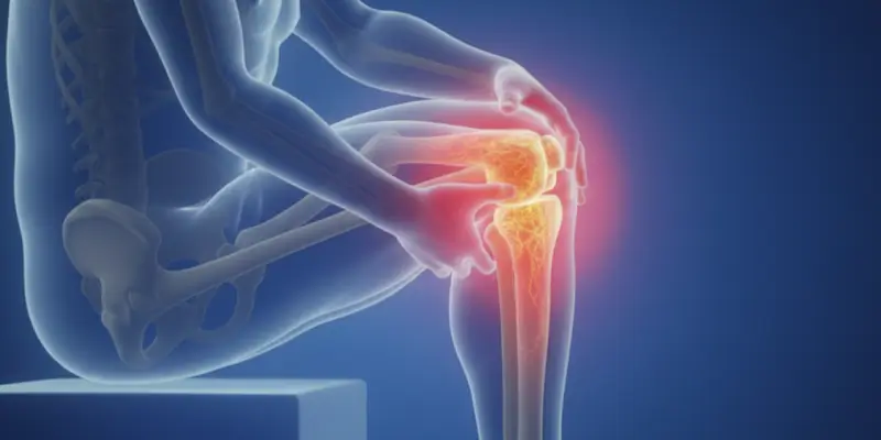
A bone infarct is a serious orthopedic disease that appears when bone tissue loses its vitality because blood flow to a certain part of the bone decreases or is cut off completely. Also called avascular necrosis or osteonecrosis in medicine, a bone infarct can over time lead to weakening of bone structure, collapse, and impairment of joint function. Especially if it is not diagnosed early, it can cause permanent damage and advanced limitation of motion.
Bones are continuously nourished by blood in order to stay healthy. This blood flow carries oxygen, nutrients, and the cells needed for repair to the bone. When this circulation is disrupted for various reasons, however, bone cells cannot receive enough nourishment and begin to die. This process is called a bone infarct. Dead bone tissue over time loses its strength, microcracks form, and bone collapse occurs especially in load-bearing regions.
A bone infarct is seen most often in the hip joint, especially in the head of the thigh bone. In addition, the bones around the knee, the shoulder, the ankle, and the wrist are among the frequently involved regions. Because these regions carry the body’s weight or move intensely, even a small disruption in blood flow can lead to serious results.
The disease can progress silently at the beginning. In the early stage the patient may feel only mild pain or stiffness. As bone structure deteriorates over time, however, pain intensifies, joint movements become limited, and in the advanced stage joint surfaces begin to collapse, which can cause permanent disability. For this reason, when bone infarct symptoms appear, evaluation without delay is of great importance.
A bone infarct is associated with many factors such as corticosteroid use, alcohol use, trauma, some blood diseases, and vessel blockages. Today, thanks to advanced imaging methods, it can be detected in the early period, and with appropriate treatment plans bone collapse can largely be prevented.
In conclusion, a bone infarct is an important bone disease that can lead to serious joint damage if it is not diagnosed in time, but that can be brought under control with early intervention. For this reason, bone and joint pain should not be taken lightly, and detailed orthopedic evaluation should always be performed especially in people who have risk factors.
Causes of a Bone Infarct
A bone infarct appears as a result of damage to or blockage of the vessels that nourish the bone. When blood flow is disrupted, bone tissue cannot receive enough oxygen and nutrients, and this leads to the development of avascular necrosis. The causes of a bone infarct directly affect both the formation of the disease and its speed of progression. For this reason, correctly identifying the underlying factors is of great importance for treatment planning.
1. Corticosteroid (Steroid) Use
Long-term or high-dose corticosteroid use is one of the most common causes of a bone infarct. Corticosteroids disrupt fat metabolism, lead to narrowing of blood vessels, and reduce blood circulation inside the bone. This increases the risk of a bone infarct in the hip and knee joints in a clear way.
2. Excessive Alcohol Use
Alcohol raises blood fats and causes clot and blockage formation inside vessels. This leads to blockage of bone vessels and increases the risk of a bone infarct. Avascular necrosis related to alcohol use is seen especially often in young and middle-aged people.
3. Trauma and Fractures
Hip dislocations, bone fractures, and serious blows can directly injure the vessels that nourish the bone. Especially after femoral neck fractures and hip dislocations, blood circulation is disrupted and a bone infarct can develop within weeks to months.
4. Blood Diseases
In some blood diseases such as sickle cell anemia, blood cells lead to blockage in vessels. This causes small vessels inside the bone to close and osteonecrosis to form.
5. Clotting Disorders
In some people, blood clots more easily than normal. These clots can block bone vessels and lead to death of bone tissue. For this reason, clotting disorders should always be investigated in recurrent bone infarct cases.
6. Chemotherapy and Radiotherapy
Some drugs used in cancer treatment and radiation can affect bone vessels and cells and set the stage for a bone infarct.
7. Autoimmune Diseases
In immune-system diseases such as lupus, both the disease itself and the drugs used affect bone vessels and increase the risk of osteonecrosis.
8. Idiopathic (Cause Unknown) Cases
In some patients, a clear cause of the bone infarct cannot be found. These cases are called idiopathic bone infarct. In these people, however, unnoticed vessel or clotting problems are generally thought to be present.
When a bone infarct is noticed early and the underlying cause is identified correctly, progression of the disease can largely be stopped. For this reason, it is very important that people who have risk factors not neglect regular orthopedic check-ups.
Which Bones Does a Bone Infarct Affect?
A bone infarct is a serious orthopedic problem that appears with death of bone tissue related to disrupted blood circulation. Avascular necrosis, its medical name, is seen more often especially in certain bone regions. The reason is that some bones have limited blood supply and are under load-bearing. In Prof. Dr. Yavuz Arıkan’s clinical approach in orthopedic oncology and bone diseases, the location of a bone infarct is accepted as one of the most important factors that determine the course of the disease and the treatment plan.
1. Thigh Bone (Femoral Head)
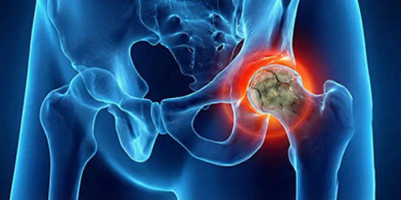
The region where a bone infarct is seen most often is the femoral head that forms the hip joint. Because this region is nourished by limited blood vessels, it is very sensitive to circulation problems.
When the femoral head is involved:
- Groin and hip pain
- Difficulty during walking
- Limping
- Joint collapse in the progressing period
can develop.
If a bone infarct in this region is not treated, it can lead to damage at a level that requires hip replacement.
2. Bones Around the Knee
A bone infarct is also frequently seen in regions such as the lower end of the thigh bone and the upper part of the shinbone that form the knee joint.
Osteonecrosis in these regions can present as:
- Knee pain
- Difficulty climbing stairs
- A locking sensation in the knee
Involvement around the knee seriously limits mobility.
3. Shoulder (Humeral Head)
The shoulder joint is also among the regions frequently affected by a bone infarct. Especially in patients who use corticosteroids, avascular necrosis can develop in the humeral head.
Symptoms:
- Shoulder pain
- Difficulty raising the arm
- Limited motion
This can over time lead to collapse and arthritis in the shoulder joint.
4. Ankle and Foot Bones
The ankle bone called the talus is prone to a bone infarct because its blood supply is limited. In this region, ankle pain, difficulty bearing weight, and swelling may be seen.
5. Wrist and Small Bones
Some small bones can also undergo osteonecrosis. Kienböck disease, in which the lunate bone is involved, is a form of wrist bone infarct.
The Difference Between a Bone Infarct and Bone Tumors
A bone infarct can sometimes be confused radiologically with a bone tumor. For this reason, differential diagnosis is of great importance. Prof. Dr. Yavuz Arıkan emphasizes that in such cases, advanced imaging and experienced orthopedic oncology evaluation are vital.
Knowing which bones a bone infarct affects is of critical importance for early diagnosis and the right treatment. Hip, knee, and shoulder pain in particular should not be neglected and should be evaluated with the necessary imaging methods.
Symptoms of a Bone Infarct
A bone infarct (avascular necrosis) is a disease that can progress silently at the beginning but over time can lead to serious bone and joint damage. With disruption of blood circulation, bone tissue loses its vitality, and as this process progresses, symptoms become gradually more obvious. According to Prof. Dr. Yavuz Arıkan’s clinical evaluations in orthopedic oncology and bone diseases, noticing bone infarct symptoms early substantially reduces the risk of joint collapse and permanent disability.
1. Deep Bone Pain That Increases with Movement
The most typical symptom of a bone infarct is bone pain of a deep, aching character.
This pain:
- May be mild at the beginning
- Becomes continuous over time
- Increases as load is applied and with movement
- May be felt as night pain
Especially in hip (femoral head), knee, and shoulder involvement, the pain spreads into the joint and keeps the patient from daily activities.
2. Limited Motion and Stiffness
As a bone infarct progresses, the joint surface is damaged. As a result, difficulty with hip or shoulder movements, trouble climbing stairs, pain when sitting down and standing up, and morning stiffness develop. These signs are important warnings that the joint surface is beginning to collapse.
3. Limping and Walking Disturbance
In a bone infarct around the hip or knee, the patient experiences limping while walking, protecting one leg when putting weight on it, and being unable to walk a distance. This can over time lead to muscle weakness and postural problems.
4. Collapse and Deformity in the Joint
As the disease progresses, dead bone tissue collapses and the joint surface deteriorates. As a result, collapse of the hip head, surface deterioration in the knee joint, and deformation in the shoulder joint can develop. This usually leads to permanent joint damage and requires surgery.
5. Swelling and Tenderness
Deep tenderness in the involved region, occasional swelling, and pain that increases when pressed may be seen. These findings show that pressure inside the bone has increased.
6. Confusion of a Bone Infarct with a Bone Tumor
A bone infarct can sometimes create appearances similar to a bone tumor on imaging methods. For this reason, differential diagnosis should be made by an experienced orthopedic oncology specialist. Prof. Dr. Yavuz Arıkan emphasizes that in such cases, MRI and advanced imaging techniques should be evaluated together to prevent incorrect diagnosis.
Bone infarct symptoms should not be taken lightly. If the diagnosis is made in the early period, bone collapse can largely be prevented and joints can be protected. For this reason, pain in the hip, knee, or shoulder that does not go away should always be evaluated by a specialist.
How Is a Bone Infarct Diagnosed?
A bone infarct is a disease whose diagnosis can be difficult especially because it does not give obvious findings on X-ray films in the early stage. For this reason, clinical examination and advanced imaging methods need to be evaluated together for an accurate diagnosis. In Prof. Dr. Yavuz Arıkan’s approach in orthopedic oncology, distinguishing a bone infarct from a bone tumor and other bone diseases is of critical importance for the patient’s future joint health.
1. Detailed Clinical Evaluation
The diagnostic process starts with the patient’s complaints. The duration and severity of pain, whether it increases with movement, whether there is night pain, corticosteroid use, alcohol history, or a history of trauma are inquired into in detail by the doctor. This information gives strong clues for a bone infarct.
2. X-ray (Plain Radiograph)
X-ray is usually the first imaging method performed. In the early stage of a bone infarct, however, the X-ray may look normal. As the disease progresses, a change in bone density, sclerotic areas, and collapse of the bone surface may be seen.
3. Magnetic Resonance Imaging (MRI)
The most sensitive method for early diagnosis of a bone infarct is MRI. Thanks to MRI, impaired circulation inside the bone, dead bone tissue, and early-stage avascular necrosis can be detected clearly.
Prof. Dr. Yavuz Arıkan emphasizes that performing MRI without delay in patients with hip, knee, and shoulder pain plays a vital role in preventing bone collapse.
4. Computed Tomography (CT)
CT is used especially for detailed evaluation of bone structure. With CT, collapse of the bone surface, microfractures, and structural deterioration are seen more clearly.
5. Bone Scintigraphy
Bone scintigraphy is a nuclear imaging method that shows bone metabolism. In regions with a bone infarct, uptake looks different because blood flow is reduced. The presence of multiple foci can be detected with this method.
6. Differential Diagnosis from a Bone Tumor
Because of imaging findings, a bone infarct can sometimes be confused with a bone tumor. For this reason, differential diagnosis is of great importance. Prof. Dr. Yavuz Arıkan recommends that in suspicious cases, MRI, CT, and clinical findings be evaluated together, and that a biopsy be performed if needed.
If a bone infarct is diagnosed in the early stage, bone collapse can be prevented and the joint can be protected. For this reason, hip, knee, or shoulder pain that does not go away should always be evaluated by an experienced orthopedic oncology specialist.
Bone Infarct Treatment Methods
Treatment of a bone infarct (avascular necrosis) is planned according to the stage of the disease, the location of the affected bone, and the patient’s general health. The main aim of treatment is to stop the progression of dead bone tissue, prevent bone collapse, and preserve joint function. In Prof. Dr. Yavuz Arıkan’s clinical approach in orthopedic oncology, joint-preserving treatments are given priority in bone infarct cases diagnosed early.
1. Follow-up and Activity Restriction
In the early stages of the disease, before bone structure has collapsed, reducing load-bearing, using crutches or support, and restricting physical activities are recommended. In this way, pressure on the damaged bone is reduced and progression can be slowed.
2. Medication
The drugs used in a bone infarct do not heal the disease directly, but they slow the process and reduce pain. Painkillers, blood thinners (to increase microcirculation), and drugs that reduce bone breakdown are used. The aim is to support circulation inside the bone and bring pain under control.
3. Reducing Pressure Inside the Bone (Core Decompression)
This method is an important surgical technique applied in the early and middle stages of a bone infarct. During the procedure, small channels are opened into the dead bone region, pressure inside the bone is lowered, and new vessel formation is encouraged. This method is especially very effective in a hip (femoral head) bone infarct and can prevent joint collapse.
4. Bone Graft and Biological Support
In advanced cases, the damaged region is cleaned and filled with healthy bone tissue or a synthetic graft. This procedure enables the bone to strengthen again. Prof. Dr. Yavuz Arıkan notes that bone graft applications are an important option for protecting the joint especially in young patients.
5. Osteotomy (Changing the Direction of the Bone)
In some patients, the angle of the bone is changed surgically to move load away from the damaged region. This method reduces pain, enables the damaged area to take less load, and prolongs joint life.
6. Joint Replacement
When a bone infarct reaches an advanced stage and the joint surface has collapsed, hip replacement, knee replacement, or shoulder replacement may be needed. This treatment provides complete elimination of pain and enables the patient to move again.
7. Differential Diagnosis from a Bone Tumor and Follow-up
Because a bone infarct can sometimes be confused with a bone tumor, accurate diagnosis before treatment is of vital importance. Prof. Dr. Yavuz Arıkan, with his orthopedic oncology experience, clarifies this distinction and prevents unnecessary or incorrect surgeries.
A bone infarct is a disease that can largely be brought under control when diagnosed in the early period. With appropriate treatment, joints can be protected, pain can be reduced, and the patient’s quality of life can be kept high.
Living with a Bone Infarct
For patients who receive a bone infarct (avascular necrosis) diagnosis, life can largely be continued in a normal way with the right treatment and informed follow-up. Because this disease can be progressive due to loss of vitality of bone tissue, however, taking some precautions in daily life and not skipping regular medical check-ups is of great importance. According to Prof. Dr. Yavuz Arıkan’s clinical approach in orthopedic oncology, people living with a bone infarct leading an active but controlled life plays a fundamental role in protecting the joints.
Daily Life and Movement
Patients with a bone infarct should not stay completely inactive, but activities that place excessive load on the affected joint should be avoided. Activities such as standing for a long time, running and jumping, and carrying heavy loads in particular can accelerate bone collapse. In contrast, swimming, a stationary bicycle, and low-impact exercises strengthen muscles and increase joint stability.
Pain Management
Pain related to a bone infarct varies according to the stage of the disease. Pain can be brought under control with regular medication use, physical therapy, and injections when needed. An increase in pain may show that the disease is progressing, so it should always be evaluated.
Nutrition and Lifestyle
To protect bone health, a diet rich in calcium and vitamin D, limiting alcohol use, and quitting smoking are very important. These measures affect bone circulation and healing capacity in a positive way.
The Importance of Regular Check-ups
A bone infarct is a chronic disease. For this reason, regular MRI and X-ray check-ups, clinical examinations, and updating the treatment plan if needed are required. Prof. Dr. Yavuz Arıkan emphasizes that a bone infarct can sometimes be confused with a bone tumor, so it is very important that imaging findings in follow-up be evaluated by a specialist eye.
Psychological and Social Life
Chronic pain and limited motion can from time to time cause low mood and anxiety. With the right treatment, however, many patients can continue working life, social activities, and daily routines. A supportive treatment plan and an informed lifestyle are the most important parts of this process.
Life with a bone infarct can be managed with accurate information, regular follow-up, and appropriate treatment. Thanks to early intervention, joints can be protected and patients can live an active life for many years.
The Difference Between a Bone Infarct and Fibrous Dysplasia
A bone infarct and fibrous dysplasia are both diseases that disrupt bone structure and can create similar appearances on imaging methods. For this reason, they can from time to time also be confused with a bone tumor. The formation mechanism, course, and treatment approaches of these two diseases, however, are completely different. In Prof. Dr. Yavuz Arıkan’s clinical practice in orthopedic oncology, making this distinction correctly is of critical importance for preventing the patient from being directed to unnecessary surgery.
Bone infarct (avascular necrosis): A bone infarct appears as a result of blockage or damage of the vessels that nourish the bone. In other words, the main problem is a blood circulation disorder. Corticosteroid use, alcohol, trauma, clotting disorders, and some diseases block bone vessels and cause the bone to die.
Fibrous dysplasia: Fibrous dysplasia, on the other hand, forms as a result of a congenital genetic mutation. In this disease, bone tissue cannot develop in a normal way and is replaced by weak fibrous tissue. There is not a blood-supply problem but a bone-formation disorder.
Fibrous dysplasia usually starts in childhood or adolescence and progresses together with bone growth. A bone infarct, on the other hand, appears more often in adult ages and over time progresses toward joint collapse.
A bone infarct most often affects the hip (femoral head), knee, shoulder, and ankle bones. Fibrous dysplasia can involve many bones such as the facial bones, the thigh, the shinbone, the ribs, and the pelvis.
Bone infarct symptoms: Deep joint pain that increases with movement, limping, limited motion, collapse of the joint surface.
Fibrous dysplasia symptoms: Bone pain, swelling, bone curvatures, easy fractures, facial and body deformities.
Both diseases create abnormal areas inside bone on X-ray, MRI, and CT. For this reason, a bone infarct can sometimes be confused with fibrous dysplasia or a bone tumor. Prof. Dr. Yavuz Arıkan emphasizes that in differential diagnosis, MRI findings and the clinical history should be evaluated together.
Bone infarct treatment is directed toward reducing pressure inside the bone, grafting, or joint replacement in the advanced stage. The aim is to protect the joint. Fibrous dysplasia treatment, on the other hand, focuses on strengthening bone, correcting deformity, and preventing fractures.
When Is a Bone Infarct Serious?
Although a bone infarct (avascular necrosis) is a disease that can be brought under control when noticed in the early stage, after certain stages it can become serious enough to lead to joint collapse and permanent loss of function. For this reason, knowing in which situations a bone infarct has reached a dangerous point is of vital importance for treatment success. In Prof. Dr. Yavuz Arıkan’s clinical approach in orthopedic oncology, the seriousness of the disease is evaluated not only according to pain but according to the structural integrity of the bone.
1. If Pain Is Continuous and Severe
If pain that was mild at the beginning over time becomes a form that continues throughout the day, wakes the person from sleep at night, and increases even with simple movements, this may show that bone tissue is gradually collapsing.
2. If Limitation of Motion Is Increasing
A clear decrease in hip, knee, or shoulder movements suggests that the joint surface is beginning to be damaged. If the patient has difficulty tying shoes, climbing stairs, or raising the arm, the bone infarct has begun to affect the joint.
3. If There Is Bone Collapse on Imaging
If collapse of the bone surface, disruption of joint congruence, or microfractures are seen on MRI or CT, the disease has reached a serious stage. At this stage, surgical treatment may be unavoidable.
4. If Load-Bearing Bones Are Involved
A bone infarct in load-bearing regions such as the hip (femoral head), knee, and ankle is much more dangerous. Collapse in these bones can in a short time lead to loss of walking, permanent limping, and the need for a prosthesis.
5. If a Bone Infarct Is Confused with a Bone Tumor
In some cases, a bone infarct can create appearances similar to a bone tumor on imaging methods. If the diagnosis is not clear, delay can lead to serious results. Prof. Dr. Yavuz Arıkan recommends that in such situations the diagnosis be confirmed with advanced imaging and biopsy if needed.
A bone infarct has reached a serious stage especially if pain is increasing, movements are becoming limited, and bone collapse is seen on imaging. At this point, specialist evaluation without losing time and an appropriate treatment plan are the only way to protect the joint.
Overall Conclusion
A bone infarct (avascular necrosis) is a progressive, serious orthopedic disease that appears when bone tissue loses its vitality as a result of disrupted blood circulation. When it affects load-bearing joints such as the hip, knee, and shoulder in particular, it directly threatens both mobility and quality of life. Clinical observations in orthopedic oncology show that when a bone infarct is not recognized in the early stage, it can create appearances that can be confused with a bone tumor.
According to the clinical approach emphasized by Prof. Dr. Yavuz Arıkan, the fundamental key to managing a bone infarct successfully is early diagnosis. With advanced imaging methods such as magnetic resonance imaging (MRI), the disease can be detected before bone collapse has developed. Thanks to protective treatments applied at this stage, procedures that reduce pressure inside the bone, and biological support methods, bone structure can be protected and serious joint damage that might develop later can be prevented.
When a bone infarct progresses, collapse of the bone surface, disruption of joint congruence, and permanent limitation of motion appear. In this situation, surgical treatments, and in some patients prosthesis applications, may be needed. With correct planning and specialist follow-up, however, even in these patients pain control can be provided and a functional life can be possible.
On the other hand, because a bone infarct can show similarity on imaging to fibrous dysplasia and some benign or malignant bone tumor types, the diagnosis should always be made by an experienced orthopedic oncology specialist. At this point, Prof. Dr. Yavuz Arıkan’s clinical approach aims for the patient to reach the most accurate diagnosis and treatment without being directed to unnecessary biopsy or operations.
In conclusion, a bone infarct is a disease that can be brought under control when caught early, and that can lead to serious joint losses when it is too late. It is of vital importance for protecting a healthy, active life that people who experience persistent pain, limited motion, or unexplained joint problems obtain specialist evaluation without delay.
Frequently Asked Questions
Frequently Asked Questions
A bone infarct (avascular necrosis) is a disease that appears when bone tissue loses its vitality because the vessels that nourish the bone are damaged. Over time the bone weakens and collapse of the joint surface can occur.
No. A bone infarct is not a bone tumor. Because it can resemble some bone tumors on imaging, however, differential diagnosis is very important. For this reason, evaluation should always be done by an experienced orthopedic oncology specialist.
A bone infarct is seen most often in the hip (femoral head), knee, shoulder, and ankle bones. Because these regions carry body weight, the disease can progress faster here.
The most common first symptom is a deep, dull joint pain that increases with movement. It may be mild at the beginning, but it intensifies over time and begins to be felt even at rest.
No. A bone infarct does not heal on its own. If untreated, it progresses and leads to bone collapse and permanent joint damage.
Yes. Long-term or high-dose corticosteroid use damages bone vessels and increases the risk of a bone infarct substantially.
A bone infarct is bone death related to a circulation problem, while fibrous dysplasia is a congenital bone development disorder. Their causes and treatments are completely different.
The most reliable diagnostic method is MRI. X-ray may be normal in the early stage. If needed, distinction from a bone tumor is made with CT and advanced imaging methods.
If untreated, the bone collapses, the joint surface deteriorates, permanent pain and loss of motion develop, and replacement surgery may be needed.
Yes. With early diagnosis and the right treatment, a large part of patients can live an active, productive life. In the clinical approach applied by Prof. Dr. Yavuz Arıkan, the aim is to preserve quality of life by protecting the bone and joint as much as possible.
 WhatsApp
  Facebook
  Twitter
  Linkedin



 +90 505 398 34 34
 doctoryavuzarikan@gmail.com
 Book Appointment / Contact