

Cartilage Transplant and Repair


What Is Cartilage and Why Does It Matter?
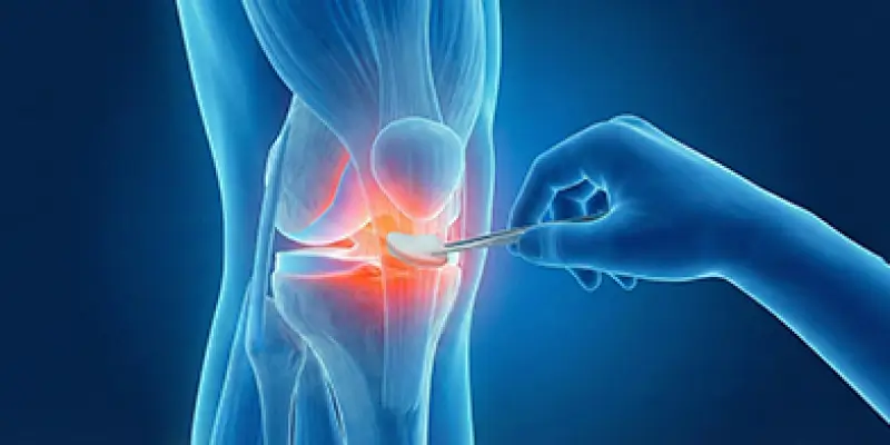
Cartilage is a smooth, slippery, flexible connective tissue that covers the ends of the bones that form joints. In mobile joints such as the knee, hip, shoulder, and ankle, it allows the bones to move comfortably without rubbing against each other. Healthy cartilage acts as a shock absorber for the joint and balances load distribution. This provides pain-free movement and prevents wear of the bones.
The most important feature of cartilage is that it contains no blood vessels or nerves. This makes it durable and smooth, but also limits its healing ability. When cartilage is damaged, its capacity to repair itself is quite low. Even a small cartilage injury can therefore progress over time to cartilage wear, joint deterioration, and osteoarthritis.
Healthy cartilage protects the joint’s range of motion and allows daily activities to be done comfortably. Basic movements such as walking, climbing stairs, squatting, and sport depend directly on cartilage quality. When the cartilage surface is disrupted, the joint surface becomes irregular, friction increases, and this causes pain, swelling, and limited motion.
Cartilage health is therefore the foundation of joint health. If cartilage loss is not noticed and treated early, it can lead in later years to serious orthopedic problems that go as far as permanent joint damage and the need for joint replacement. Modern treatments such as cartilage repair and cartilage transplant aim to protect the joint and continue natural movement for many years.
What Is Cartilage Damage?
Cartilage damage is wear, cracking, or complete loss of the smooth cartilage layer covering the joint surface. It is seen most often in the knee, hip, and ankle. Healthy cartilage allows the bones to move without rubbing; when cartilage damage develops, the joint surface is disrupted, friction increases, and pain appears.
Because cartilage contains no blood vessels, its capacity to repair itself when damaged is extremely limited. Even a small cartilage injury can therefore progress over time to cartilage wear, disruption of the joint surface, and osteoarthritis. If untreated, cartilage loss becomes difficult to reverse.
Cartilage damage usually appears in two main ways. The first is traumatic cartilage damage, which occurs after sudden impacts such as sports injuries, falls, and twisting of the knee. The second is degenerative cartilage damage, which is gradual wear of cartilage over many years of load-bearing, related to age. Obesity, knee deformity, and incorrect loading can also speed this process.
In the damaged cartilage area the joint surface becomes rough. This leads to complaints such as a catching sensation, pain, swelling, and stiffness during movement. Over time bone surfaces are exposed and bone begins to contact bone. This can cause both severe pain and permanent joint deformities.
Cartilage damage is therefore an important orthopedic problem that should be diagnosed and treated early. With modern treatments such as cartilage repair and cartilage transplant, damaged joint surfaces can be repaired and the natural joint structure protected for a long time.
What Are the Symptoms of Cartilage Damage?
Symptoms of cartilage damage can vary with the joint involved, the size of the damage, and the degree of progression. Because the most frequently affected areas are the knee, hip, and ankle, complaints usually concentrate in these joints. Because cartilage does not contain pain nerves, mild complaints may be seen early; as damage progresses, symptoms become more obvious.
The most common symptom is joint pain. Knee or hip pain appears especially when climbing stairs, squatting, standing for a long time, or playing sport. Pain that increases with movement at the beginning can later be felt even at rest.
Another important symptom is swelling. Damaged cartilage can cause an inflammatory reaction inside the joint and lead to an increase in joint fluid. Visible swelling can form in the knee or ankle.
Catching, locking, and a rubbing sensation appear when the cartilage surface is disrupted. Rough surfaces rub against each other during movement and this can be felt as a “click” or “cracking” sound.
Limited motion is also an important sign of cartilage damage. The joint stiffens, cannot fully open and close, and daily movements become harder. This is more obvious especially in the morning or after remaining still for a long time.
In advanced cartilage loss, loss of strength and a sense of unsteadiness can develop. Because the knee or hip cannot provide enough support, a sense of insecurity and fear of falling can appear while walking.
If these symptoms are not assessed in time, cartilage damage can progress to osteoarthritis. People with long-lasting pain, swelling, and limited motion in the knee, hip, or ankle should therefore see an orthopedic specialist.
How Is Cartilage Damage Diagnosed?
Correct diagnosis of cartilage damage is extremely important for treatment success. Because cartilage has a low capacity to repair itself, noticing it early protects the joint. Diagnosis is made both by assessing the patient’s complaints and by advanced imaging.
In the first stage the orthopedic specialist asks in detail about complaints such as pain, swelling, a catching sensation, and limited motion. A joint examination is then done. Range of motion of the knee, hip, or ankle, tender points, whether there is locking, and stability are assessed. This examination provides important clues about the presence of cartilage damage.
Among imaging methods, the most valuable investigation is MRI (magnetic resonance imaging). MRI shows in detail the thickness of the cartilage layer, tears, wear areas, and underlying bone changes. Knee cartilage damage and hip cartilage problems in particular can be detected with high accuracy by MRI.
X-ray does not show cartilage directly, but it provides information about indirect findings such as narrowing of the joint space, disruption of the bone surface, and osteoarthritis. In patients with advanced cartilage loss, X-ray is an important complementary investigation.
In some cases arthroscopy is used to confirm the diagnosis. A small camera is introduced into the joint and the cartilage surface is seen directly. Arthroscopy offers the possibility of both diagnosis and treatment at the same time.
With a correct diagnosis, the degree of cartilage damage is determined and cartilage repair, cartilage transplant, or other biological treatments can be planned accordingly. Cartilage problems diagnosed early can be controlled before osteoarthritis develops.
What Is Cartilage Repair?
Cartilage repair is the general name for modern treatments that aim to rebuild or improve damaged or lost cartilage on the joint surface. The main aim of these treatments is to make the joint surface smooth again, reduce pain, increase mobility, and prevent osteoarthritis.
Because cartilage contains no blood vessels, its capacity to heal by natural means is very low. Damage after trauma, sports injury, or age-related cartilage wear can therefore progress over time. Cartilage-repair methods activate the body’s healing mechanisms so that the damaged area is covered again with a healthy surface.
Cartilage repair is usually applied by arthroscopic (closed) methods. The damaged area is cleaned and the bone surface underneath is stimulated so that a new cartilage-like tissue forms. One of the most frequently used methods is the microfracture technique. Small holes are opened in the bone surface so that stem cells reach the damaged area and a new cartilage layer forms.
In more advanced cartilage damage, real cartilage tissue can be placed with methods such as cartilage cell transplant or mosaicplasty. These methods are preferred especially in young and active patients with knee cartilage damage and ankle cartilage problems.
With cartilage repair, patients can largely be freed from complaints such as knee pain, a catching sensation, and limited motion. When applied in the right patient and with the appropriate technique, cartilage repair is an effective treatment option that delays the need for joint replacement and protects the natural joint.
What Is Cartilage Transplant?
Cartilage transplant is the placement of new, healthy cartilage cells or cartilage tissue in place of cartilage that is severely damaged or completely lost on the joint surface. This method is applied especially in patients with limited but deep cartilage damage in the knee, hip, and ankle. The aim is to make the joint surface smooth again, reduce pain, and restore the joint’s natural function.
Because cartilage cannot renew itself, simple treatments are not enough in advanced cartilage loss. This is where cartilage transplant comes in. The patient’s own cartilage cells or donor-derived cartilage tissue is placed in the damaged area. A structure closest to real cartilage tissue is thus obtained.
Especially in young, active patients without advanced osteoarthritis, cartilage transplant can delay major operations such as knee replacement for many years. For athletes and people who lead an active life, protecting the joint’s natural structure is a great advantage.
Cartilage Transplant and Repair Methods
Different cartilage repair and transplant methods are applied according to the size and depth of the cartilage damage and the patient’s age. The most frequently used methods are:
Microfracture
The damaged cartilage area is cleaned and small holes are opened in the bone surface underneath. Stem cells coming from these holes form a new cartilage-like tissue. It is effective in small and medium-sized cartilage damage.
Mosaicplasty (OATS)
Small cartilage–bone pieces taken from a healthy area of the patient’s own joint are placed in the damaged area. Repair is thus provided with real cartilage tissue. It is preferred in medium-sized cartilage loss.
Cartilage Cell Transplant (ACI – MACI)
In this advanced technique, the patient’s own cartilage cells are multiplied in the laboratory and then transplanted to the damaged area. It is one of the most successful methods in large and deep cartilage loss.
Stem-Cell-Supported Cartilage Repair
Stem cells obtained from bone marrow or fat tissue are applied to the damaged area to support cartilage regeneration.
With these modern cartilage-repair and cartilage-transplant methods, the joint surface can be made healthy again, pain decreases, and the development of osteoarthritis can be slowed to a large extent.
Who Is Cartilage Transplant Applied To?
Cartilage transplant is an advanced treatment applied in patients with limited but deep cartilage loss on the joint surface whose overall joint structure is still protected. This treatment provides a great advantage especially for people who want to protect their natural joint and continue an active life.
The most suitable candidates for cartilage transplant are usually patients under 50 with localized cartilage damage in the knee, ankle, or hip. This method is quite successful especially in cartilage loss related to sports injuries, trauma, or old ligament injuries.
Patients without advanced osteoarthritis form the most suitable group for cartilage transplant. If there is widespread cartilage wear and bone deformity throughout the joint, cartilage transplant is not enough and larger surgeries such as joint replacement come onto the agenda.
Cartilage transplant is also an ideal option for:
- Those with kneecap (patella) cartilage damage
- Cartilage problems that develop after ACL surgery
- Patients with ankle cartilage lesions
- Individuals whose joint stability has been restored
Excess weight, active infection, advanced joint disorder, and smoking, however, can adversely affect treatment success. Patient selection therefore plays a critical role in the success of cartilage transplant.
When applied in the right patient, cartilage transplant reduces pain, increases mobility, and can delay the need for joint replacement for many years.
How Is Cartilage Transplant Surgery Performed?
Cartilage transplant surgery is an advanced procedure that aims to cover the damaged joint surface again with a healthy cartilage layer. This operation is usually applied in the knee, ankle, and in some cases the hip. The aim is to eliminate pain, catching, and limited motion related to cartilage loss while protecting the joint’s natural structure.
Before surgery, the location, depth, and size of the cartilage damage are assessed in detail with MRI. Which cartilage-transplant method will be used is thus determined.
Surgery is usually done arthroscopically (closed) or, when needed, through a small open incision. In the first stage the damaged cartilage tissue is completely cleaned and the bone surface underneath is prepared. Then, according to the method chosen:
- If mosaicplasty is applied, cartilage–bone cylinders taken from a healthy area of the patient’s own joint are placed in the damaged area.
- If cartilage cell transplant (ACI/MACI) is to be done, cartilage cells previously multiplied in the laboratory are applied to the damaged area on a special membrane or carrier.
- In biologically supported transplants, repair is supported with stem cells or growth factors.
After transplant, the newly placed cartilage tissue is secured and the joint surface is made smooth again. Surgery usually lasts 1–2 hours and most patients are discharged within a few days.
Maturation of the new cartilage tissue after cartilage transplant takes time. Controlled loading and a physiotherapy program are therefore critical for treatment success. With the correct surgical technique and appropriate rehabilitation, cartilage transplant can protect joint health for many years.
The Recovery Process After Surgery
The recovery process after cartilage transplant and cartilage repair is one of the most important stages that directly affect surgical success. Because attachment of the newly placed cartilage tissue to bone and its maturation take time, the patient’s full compliance with the doctor’s recommendations in this period matters a great deal.
In the first days after surgery, swelling and mild pain in the joint may be seen. This is normal and is controlled with medication. In patients who have had knee or ankle cartilage transplant, walking with crutches is usually recommended. Full load is not given to the operated joint for the first 4–6 weeks. The new cartilage tissue thus attaches to bone without being harmed.
Physiotherapy usually starts a short time after surgery. At the beginning, joint stiffness is prevented with passive movements; later, muscle strengthening and balance exercises are added. After knee cartilage surgery, strengthening of the muscles is very important for joint stability.
Full maturation of the new cartilage tissue can take about 6–12 months. During this period, high-impact sports, sudden turns, and jumping are not recommended. Activities that do not load the joint, such as walking, swimming, and cycling, support recovery.
With the right rehabilitation and regular follow-up, joint pain decreases after cartilage transplant, mobility increases, and return to daily life takes place in a healthy way. This process is critical for the lasting effect of cartilage treatment.
Is Cartilage Transplant Lasting?
Cartilage transplant, when applied in the right patient and with the appropriate technique, is an advanced treatment that can provide lasting results for many years. The main aim of this procedure is to cover the damaged joint surface again with healthy, durable cartilage tissue and protect the joint’s natural structure. Pain thus decreases, mobility increases, and the progression of osteoarthritis is slowed to a large extent.
The lasting effect of cartilage transplant depends most on the stage of disease, the size of the damage, and the patient’s lifestyle. If cartilage loss is in a limited area and there is no widespread osteoarthritis throughout the joint, the transplanted cartilage tissue attaches strongly to bone and can function healthily for years.
Especially in young, active patients whose joint structure is not disrupted, methods such as cartilage cell transplant (ACI) and mosaicplasty can delay the need for joint replacement for a long time. Patients can continue their lives without pain with their own natural joints.
Excess weight, smoking, activities that overload the joint, and inadequate rehabilitation, however, can shorten the life of the transplanted cartilage. In patients with advanced osteoarthritis, cartilage transplant may not offer a lasting solution and other treatments may be needed over time.
With the right patient selection, successful surgical technique, and regular follow-up, cartilage transplant offers a long-term, effective solution in knee, hip, and ankle cartilage damage.
The Difference Between Cartilage Transplant and Joint Replacement
Cartilage transplant and joint replacement are two different surgical approaches used in the treatment of cartilage loss that causes joint pain and limited motion. The main difference between these two methods is whether the joint is protected.
Cartilage transplant is a treatment that protects the joint’s natural structure. Damaged cartilage tissue is repaired or new cartilage tissue is placed in its place. The aim is to make the joint surface smooth again so that the bones move in a natural way. This method is preferred especially in young, active patients with limited cartilage damage. With cartilage transplant, the patient continues life with their own knee, hip, or ankle joint.
Joint replacement is the complete removal of the damaged joint and placement of an artificial joint. This method is applied when the joint surface is severely disrupted, bone collapse has occurred, or advanced osteoarthritis has developed. Joint replacement largely eliminates pain, but the joint’s natural structure is no longer present.
Cartilage transplant protects joint biomechanics and the sense of movement is more natural. In joint replacement, movement takes place on an artificial surface. Implants also have a limited lifespan, and in young patients in particular another operation may be needed in later years.
In early-stage cartilage damage, cartilage repair and cartilage transplant are therefore much more advantageous than joint replacement. Joint replacement is usually preferred as a last resort when all other treatment options have been exhausted.
Advantages of Cartilage Transplant
Cartilage transplant is one of the most advanced, joint-preserving methods used in the treatment of cartilage loss on the joint surface. Especially in knee, hip, and ankle cartilage damage, it offers important advantages in providing long-term recovery while protecting the natural joint.
The greatest advantage of cartilage transplant is that it protects the joint’s natural structure. Unlike joint-replacement operations, the joint is not completely replaced; only the damaged area is repaired. Joint biomechanics are thus not disrupted and the sense of movement remains natural.
Another important advantage is a clear reduction in pain. When the damaged cartilage area is repaired, rubbing of the bones against each other is eliminated. This largely reduces chronic pain in the knee, hip, or ankle.
Cartilage transplant increases mobility. Because the joint surface becomes smooth again, the patient can walk more comfortably, climb stairs, and do daily activities without pain. For people who play sport or lead an active life, this is a great gain.
Another important advantage is that it delays osteoarthritis. When cartilage loss is repaired early, bone deformities and advanced osteoarthritis that could form later can be prevented. This can postpone the need for hip or knee replacement for many years.
Cartilage transplant is also an ideal solution for young and middle-aged patients. Given that implants have a limited lifespan, this method that protects the natural joint provides a great long-term advantage.
For all these reasons, cartilage transplant is a modern treatment option that, in suitable patients, protects joint health, improves quality of life, and offers lasting recovery.
Risks of Cartilage Transplant
Cartilage transplant is an advanced procedure that aims to protect the joint and rebuild natural cartilage tissue. Although it is generally a safe method, like every surgical procedure it carries certain risks. These risks can vary with the patient’s health, surgical technique, and the cartilage-transplant method applied.
1. Infection Risk
As with every surgical procedure, there is a risk of infection in cartilage transplant surgery. Infection inside the joint is rare, but if it occurs treatment can take a long time and joint health can be adversely affected.
2. Risk That the New Cartilage Tissue Does Not Take
In cartilage transplant, attachment of the transplanted cells or cartilage pieces to bone is very important. If adequate attachment is not achieved, new cartilage tissue may not develop and surgical success may fall. This can be seen especially in heavy cartilage loss or older patients.
3. Pain and Swelling
Temporary pain and swelling in the joint after surgery are common. This usually decreases within a few weeks. In some patients, however, pain may last and restrict joint movement.
4. Joint Stiffness and Limited Motion
If appropriate rehabilitation is not done after surgery, joint stiffness and limited motion can develop. Physiotherapy and controlled exercises are therefore critical for reducing risks.
5. Bleeding and Vessel Injury
Rarely, bleeding or vessel injury around the surgical field can be seen in cartilage transplant surgery. Such complications are usually controlled during surgery.
6. Repeat Damage and Long-Term Risks
Excessive loading after surgery, smoking, or joint trauma can lead to repeat damage of the transplanted cartilage. Lifestyle changes after surgery and compliance with the doctor’s recommendations are therefore very important.
Conclusion
Cartilage tissue is the basic building block of joint health and protects joint function by allowing the bones to move without friction. Trauma, sports injuries, age-related degeneration, or hereditary factors, however, can lead to cartilage damage. If cartilage damage is not treated, complaints such as pain, swelling, joint stiffness, and limited motion appear, and in the later period it can cause osteoarthritis.
Cartilage repair and cartilage transplant are modern, effective surgical methods used to repair or rebuild damaged cartilage on the joint surface. Microfracture, mosaicplasty (OATS), autologous cartilage cell transplant (ACI/MACI), and stem-cell-supported repair techniques are chosen according to the size of the damage and the patient’s age. With these methods the joint protects its natural structure, pain decreases, mobility increases, and the need for joint replacement can be postponed for many years.
Cartilage transplant is preferred especially in young, active patients whose joint structure is not disrupted. During surgery, damaged cartilage is cleaned and healthy cartilage tissue or multiplied cartilage cells are placed in its place. These procedures, done arthroscopically or through a small incision, allow the patient to be discharged quickly and return to daily life in a short time with a controlled rehabilitation process.
The recovery process after surgery is critical for the success of cartilage transplant. Load restriction is applied in the first weeks, and joint mobility and muscle strengthening are provided with physiotherapy. Maturation of the new cartilage tissue usually takes 6–12 months, and in this process an appropriate lifestyle and avoiding smoking and excessive loading are important.
With the right patient selection, appropriate surgical technique, and regular follow-up, cartilage transplant offers lasting results for many years. Its difference from joint-replacement operations is that it protects the joint’s natural structure and reduces pain without disrupting biomechanics. Although risks are rare, situations such as infection, failure of the tissue to take, or joint stiffness can be seen; an experienced surgeon and careful follow-up are therefore essential.
In conclusion, cartilage transplant and repair are a modern, safe, and effective treatment option for patients who want to protect joint health, increase mobility, and reduce pain. When applied early, it delays osteoarthritis, postpones the need for joint replacement, and significantly raises quality of life.
Frequently Asked Questions
Frequently Asked Questions
Cartilage transplant is the placement of healthy cartilage cells or tissue pieces in place of damaged cartilage on the joint surface. The aim is to reduce pain and increase mobility while protecting the joint’s natural structure.
Cartilage transplant is generally suitable for patients under 50 with limited cartilage damage, without advanced osteoarthritis, who lead an active life. Young athletes and people with knee, hip, or ankle damage are the most suitable candidates.
Surgery is usually done arthroscopically. Damaged cartilage is cleaned and the bone surface underneath is prepared. Depending on the method chosen, healthy cartilage cells or cartilage–bone pieces are placed in the damaged area. The procedure usually lasts 1–2 hours.
Load restriction is applied for the first 4–6 weeks after surgery and crutches are used. Physiotherapy provides joint mobility and muscle strengthening. Maturation of the new cartilage tissue can take 6–12 months.
With the right patient selection and appropriate surgical technique, cartilage transplant can provide lasting results for many years. The transplanted cartilage tissue attaches to bone and protects joint function for a long time.
Cartilage transplant protects the joint and provides natural movement. Joint replacement completely replaces the joint. In young and active patients, cartilage transplant is preferred first; joint replacement is usually used in patients with advanced osteoarthritis or joint deformity.
It protects the joint’s natural structure, reduces pain, increases mobility, delays osteoarthritis, and postpones the need for joint replacement for many years.
Rare risks such as infection, failure of the tissue to take, joint stiffness, or temporary pain can occur. With an experienced surgeon and appropriate rehabilitation, these risks are minimized.
Controlled activities are recommended for the first 6–12 months. Low-impact sports such as walking, cycling, and swimming support recovery. Load-bearing activities that require sudden movement are started gradually under medical supervision after surgery.
Cartilage transplant is applied most often in the knee, hip, and ankle. It can also be used in suitable patients in joints such as the shoulder and elbow, but the field of application is limited and specialist assessment is required.
 WhatsApp
  Facebook
  Twitter
  Linkedin





 +90 505 398 34 34
 doctoryavuzarikan@gmail.com
 Book Appointment / Contact