

What Is Cervical Disc Herniation? Symptoms and Treatment


Why Is Cervical Disc Herniation So Common?
Cervical disc herniation is one of the most common spinal problems of modern life. In Prof. Dr. Yavuz Arıkan’s clinical approach in orthopedic oncology, longer desk work and widespread smartphone and tablet use have increased load on the cervical spine. Staying bent forward for long periods constantly strains the neck muscles, contributes to early disc degeneration, and over time sets the stage for herniation. Cervical disc herniation is therefore no longer seen only as a disease of older age; it is increasingly common in young adults.
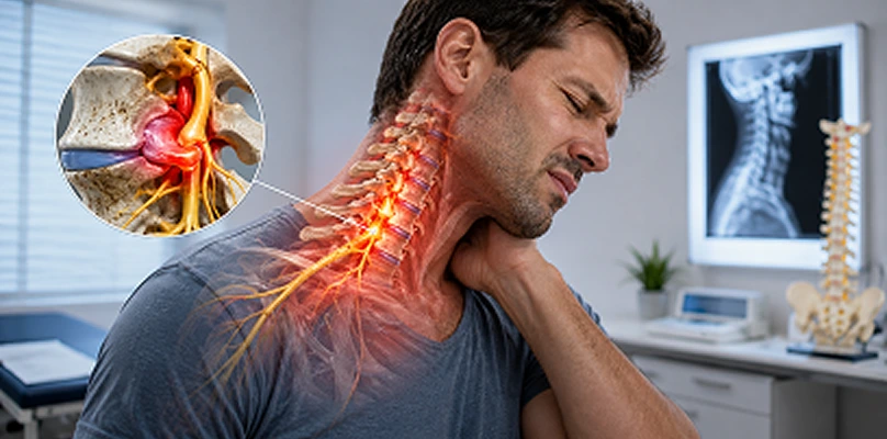
Cervical disc herniation occurs when the disc between the vertebrae is damaged, the inner material (nucleus pulposus) bulges outward, and pressure is placed on a nerve root or the spinal cord. This pressure can cause pain and neurologic findings. The neck is a critical region where the spinal cord passes and the nerves to the upper limbs exit, so pathology here can have serious consequences. In every patient who presents with neck pain, cervical disc disease should be considered, not only simple muscle spasm.
Cervical Spine Anatomy and the Role of the Disc
The cervical spine consists of seven vertebrae (C1–C7). C1 and C2 have a special structure for head motion and rotation, while C3–C7 mainly bear load and provide stability. The discs between the vertebrae give the spine flexibility and motion. The outer part of the disc is the fibrous annulus fibrosus; the inner part is the gel-like nucleus pulposus. This structure allows motion and distributes load.
Cervical discs herniate most often at C5–C6 and C6–C7, because these levels carry the greatest motion and mechanical load. Disc degeneration and herniation usually progress slowly and accumulate over years into a clear clinical picture.
How Cervical Disc Herniation Develops (Pathophysiology)
Cervical disc herniation usually comes from a degenerative process over years, not from a single sudden trauma. With age, discs lose water content and elasticity. The outer ring (annulus fibrosus) weakens from repeated microtrauma and may tear. Through these tears, the nucleus pulposus can bulge outward.
At this stage a “protrusion,” or bulge, may form. Later the annulus tears and disc material herniates more clearly. If this material presses on a nerve root or the spinal cord, clinical symptoms appear. In some patients a small herniation causes severe pain because of inflammation and chemical irritation of the nerve root. In others a large herniation causes milder symptoms, depending on its direction and relationship to nerve tissue.
Risk Factors: Who Is More Affected?
Many factors play a role. The most common include:
- Prolonged desk work: A screen below eye level increases forward neck tilt.
- Technology use: Bending the head forward with a phone or tablet increases pressure on cervical discs.
- Smoking: Impairs disc nutrition and speeds degeneration.
- Genetic predisposition: Risk is higher in people with a family history of disc disease.
- Occupation: Drivers, dentists, and workers who stand for long periods are at higher risk.
- Obesity: Excess weight increases spinal load and speeds disc degeneration.
- Trauma: Traffic accidents, falls, or sudden strain can damage the disc.
Some of these factors can be controlled; genetic predisposition cannot. Managing controllable risks is therefore important for slowing development and progression.
Clinical Symptoms of Cervical Disc Herniation
Symptoms depend on the level of the herniation and the degree of nerve compression. The most common complaint is neck pain. Pain often starts at the nape and radiates to the shoulder and arm. Staying in one position, working at a computer, or sudden neck movements can increase pain.
Pain is often “radicular,” felt in the dermatome of the affected nerve root. It may feel like burning, stabbing, or electric shock. Numbness and tingling are also common and help identify the affected root. For example, C6 compression often causes numbness in the thumb and index finger, C7 in the middle finger, and C8 in the ring and little fingers.
Reduced muscle strength, weaker grip, and impaired fine motor skills appear in more advanced cases and can seriously affect daily life. When spinal cord compression is present, the picture is more severe: gait disturbance, loss of balance, weakness in the hands, and rarely problems with bladder control. These symptoms require urgent evaluation.
Clinical Evaluation in Diagnosis
Diagnosis starts with a detailed history and physical examination. The character, severity, and radiation of pain, which movements increase it, and its effect on daily life are asked. Range of motion, muscle tone, reflexes, sensation, and strength are assessed. Special maneuvers such as the Spurling test help detect nerve-root compression.
Neurologic examination is the most critical element in diagnosis and planning. Weakness, reflex loss, or sensory change show which level is affected and how much. Patients with neck pain therefore need assessment of neurologic function, not only pain.
Imaging: Which Test Does What?
Imaging is needed to confirm clinical findings and plan treatment. Common methods include:
- X-ray: Shows alignment, disc-space height, and degenerative change. The herniation itself is not seen, but reduced disc height and spinal stability can be assessed.
- Magnetic resonance imaging (MRI): The gold standard. Disc structure, nerve roots, and the spinal cord are evaluated in detail. MRI shows the size of the herniation and its effect on nerve tissue.
- Computed tomography (CT): More useful for bone. It is used especially to assess spinal canal stenosis.
- Electromyography (EMG): Helps determine the degree of nerve injury and which root is affected. EMG is important when clinical findings and MRI do not match.
These studies also help determine the treatment course and possible need for surgery.
Basic Principles of Treatment
The main goals are:
- Controlling pain
- Reducing nerve compression
- Preserving function
- Returning to daily activities
- Reducing the risk of recurrence
The plan is individualized according to age, duration of symptoms, pain severity, neurologic findings, imaging, and lifestyle. Most patients improve with nonsurgical care; in some cases surgery is needed.
Conservative (Nonsurgical) Treatment
The first step is usually conservative care. The aims are to reduce pain, control inflammation, and preserve neck function.
1. Medication
Medication is adjusted to pain severity and general health. Common groups include:
- Painkillers (paracetamol/acetaminophen): Preferred for mild to moderate pain.
- Nonsteroidal anti-inflammatory drugs (NSAIDs): Reduce inflammation and control pain.
- Muscle relaxants: May be used to reduce spasm and stiffness.
- Nerve-pain (neuralgia) medicines: When nerve-root irritation causes severe pain, drugs such as gabapentin or pregabalin may help.
The lowest effective dose should be used and side effects monitored. With NSAIDs, stomach, kidney, and cardiovascular risks must be considered.
2. Physical Therapy and Exercise
Physical therapy is a core part of treatment. The program is personalized. Goals include:
- Strengthening neck muscles
- Improving posture
- Stabilizing the shoulder and upper-back muscles
- Preserving neck range of motion
- Pain control
Exercise usually starts slowly and with controlled, pain-free movements. The most effective exercises include isometric neck work, scapular stabilization, and programs that strengthen the back muscles. Regular practice reduces pain and lowers recurrence risk.
3. Lifestyle Changes and Ergonomics
Lifestyle changes are among the most critical factors for success. Important adjustments include:
- The computer screen should be at eye level.
- Elbows should be supported at about 90 degrees when using keyboard and mouse.
- The head should not be bent forward when using a phone.
- Avoid staying in one position; take short breaks every 30–45 minutes.
- Bed and pillow choice should support neck physiology.
These changes speed recovery and reduce recurrence.
Injection and Interventional Treatments
In some patients conservative care is not enough. When severe radicular pain is due to nerve-root compression, injections may be considered. They can control pain so the patient can take part more effectively in physical therapy.
- Epidural steroid injection: Reduces inflammation around the nerve root and eases pain.
- Nerve-root blocks: Injection around the targeted root to control pain.
- Facet joint blocks: Used when neck pain comes from the facet joints.
Effect varies from person to person. Success depends on correct patient selection and technique. These procedures are usually combined with conservative care.
Surgical Treatment: When Is It Needed?
Surgery is considered when specific indications are present. The priority is to relieve nerve compression and prevent neurologic loss. Indications include:
- Progressive neurologic loss (increasing weakness, reflex loss)
- Spinal cord compression and myelopathy (gait disturbance, balance problems, reduced hand skill)
- Severe pain that does not respond to conservative care
- Pain that seriously impairs quality of life
The most common techniques include:
- Anterior cervical discectomy and fusion (ACDF): The disc is removed and the vertebrae are fused.
- Disc replacement (total disc arthroplasty): The disc is removed and a mobile prosthesis is placed.
- Posterior approach: May be preferred in multilevel herniation or spinal cord compression.
Choice of method depends on age, level of herniation, spinal stability, and general health. Microsurgical techniques have made recovery faster and more comfortable.
Rehabilitation After Surgery
The postoperative period determines long-term function. Early on, pain control, wound care, and mobilization are the priority. Physical therapy usually starts a few weeks after surgery and progresses according to the patient’s condition.
Main goals of rehabilitation:
- Preserving neck range of motion
- Regaining muscle strength
- Improving posture
- Returning to daily activities
Patient education is also important. Attention to ergonomics, avoiding heavy lifting, and regular exercise reduce recurrence.
What Happens If Cervical Disc Herniation Is Not Treated?
If untreated, pain and neurologic findings may progress. Ongoing nerve-root compression can reduce muscle strength and cause permanent nerve injury. Spinal cord compression can lead to gait disturbance, loss of balance, and reduced hand skill. These can substantially reduce quality of life and cause damage that is hard to reverse.
Chronic pain also changes lifestyle; inactivity, muscle weakness, and psychological problems may develop. Neck pain and symptoms radiating into the arm should not be ignored; specialist evaluation should be sought early.
Prevention and Lifestyle Advice
The most effective prevention methods are ergonomics and regular exercise. During computer and phone use, the screen should be at eye level and prolonged static posture avoided. Regular walking, swimming, and exercises that strengthen the shoulder and back muscles support cervical spine health. Pillow and mattress choice also matter; orthopedic pillows that support neck physiology should be preferred.
Smoking should be stopped and weight controlled. Obesity increases spinal load and speeds disc degeneration. Lifestyle change is therefore a cornerstone of treatment.
Conclusion
Cervical disc herniation is a spinal condition that can largely be controlled with correct diagnosis and appropriate treatment. With early diagnosis and an individualized plan, most patients recover with conservative methods and do not need surgery. When progressive neurologic findings are present, surgery can be life-saving and reduce the risk of permanent damage. Prof. Dr. Yavuz Arıkan emphasizes that people with pain radiating from the neck into the arm should seek specialist evaluation without delay.
Frequently Asked Questions

Specialist Opinion
Cervical disc herniation is a spinal condition that can largely be controlled with correct diagnosis and appropriate treatment. With early diagnosis and an individualized plan, most patients recover with conservative methods and do not need surgery. When progressive neurologic findings are present, surgery can be life-saving and reduce the risk of permanent damage. Prof. Dr. Yavuz Arıkan emphasizes that people with pain radiating from the neck into the arm should seek specialist evaluation without delay.
Contact us to have your symptoms assessed and to learn about treatment options that may be right for you. Get in touch .
Frequently Asked Questions
No. Disc degeneration increases with age, but prolonged poor posture, trauma, and heavy technology use mean it can also occur in younger people.
The disc may not return fully to normal, but symptoms can often be controlled with appropriate treatment and lifestyle changes. In some patients the herniation may recede over time.
Neck pain, burning or tingling radiating into the arm, muscle weakness, and reflex changes are important clues. Definitive diagnosis is made with MRI.
Isometric neck muscle work, scapular stabilization exercises, and programs that strengthen the upper back muscles are often recommended.
Conservative treatment is enough for most patients. Surgery is considered for progressive neurologic loss or severe pain that does not respond to conservative care.
 WhatsApp
  Facebook
  Twitter
  Linkedin



 +90 505 398 34 34
 doctoryavuzarikan@gmail.com
 Book Appointment / Contact