

What Is Osteoid Osteoma? Symptoms, Diagnosis, and Treatment


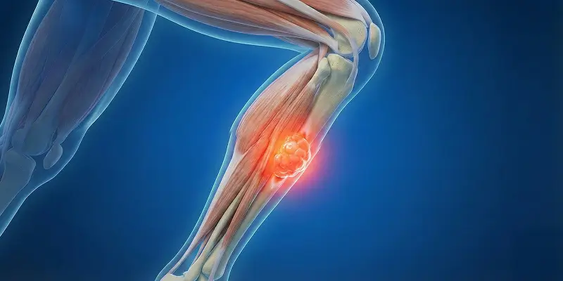
Osteoid osteoma is a benign bone tumor seen especially in children and young adults. It usually appears in long bones and is known for severe, characteristic pain despite its small size. It is one of the most common benign bone lesions and typically presents with bone pain that increases at night.
The tumor forms when osteoblasts, the bone-forming cells, become overactive. This activity creates a small but dense tumor focus (nidus) inside the bone. Osteoid osteoma is usually 1–2 cm in diameter, but it causes marked hardening and inflammation in the surrounding bone.
What Is Osteoid Osteoma?
Osteoid osteoma is a small tumor of osteoid tissue that sits in the bone cortex or metaphysis. At its center is an active area called the “nidus.” The nidus produces large amounts of prostaglandins, substances that cause pain. That is why osteoid osteoma can cause very severe pain despite its small size.
Unlike malignant bone cancers, it does not spread through the body (it does not metastasize). If untreated, however, it can lead to bone deformity, muscle weakness, and loss of function.
Who Gets It?
Osteoid osteoma is most common in:
- People aged 10–25
- Males (2–3 times more often than females)
- Active young people who play sports
The bones most often affected are:
- Femur (thigh bone)
- Tibia (shinbone)
- Spine
- Pelvis
- Bones of the feet and hands
Osteoid Osteoma Symptoms
The most characteristic finding is bone pain that increases at night. This pain differs from ordinary muscle or joint pain.
The most common symptoms are:
- Severe pain that increases at night
- Bone pain that continues even at rest
- Temporary relief with NSAIDs (aspirin, ibuprofen, and similar drugs)
- Tenderness in the affected area
- Swelling or muscle spasm in some patients
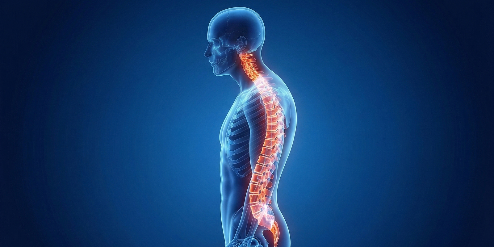
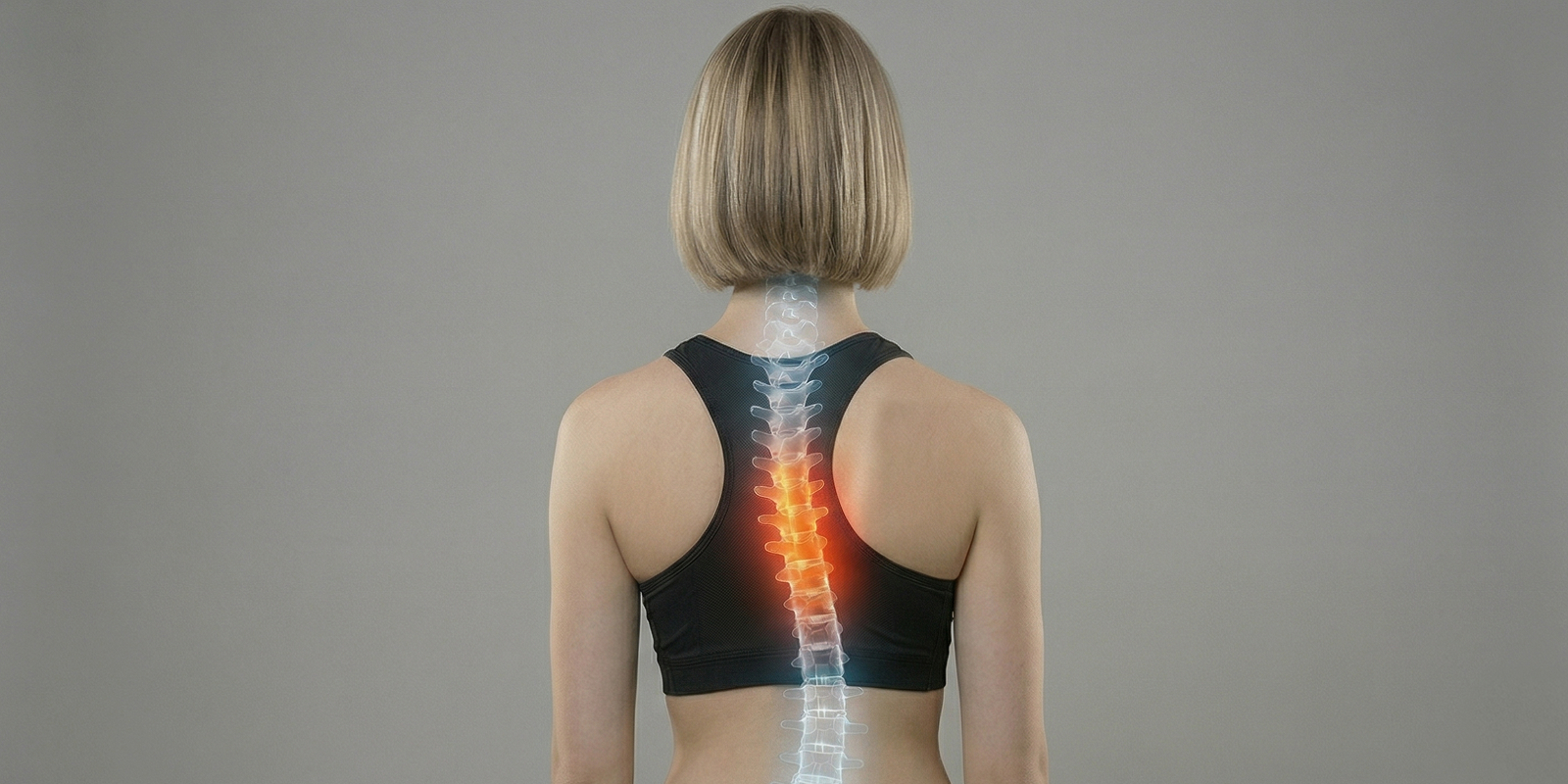
- In the spine, scoliosis or postural change
Pain may be mistaken for months or even years as “growing pains” or a sports injury.
How Is Osteoid Osteoma Diagnosed?
Diagnosis is based on clinical findings and advanced imaging.
X-ray
This is the first-line study. In the early period, the nidus is not always clearly visible.
CT (Computed Tomography)
This is the gold standard. CT shows the nidus at millimeter scale and confirms the diagnosis.
MRI (Magnetic Resonance Imaging)
MRI shows edema and inflammation in surrounding tissues. Used alone, it can miss the nidus, so it is interpreted together with CT.
At specialist centers, CT-supported evaluation by physicians experienced in orthopedic oncology, such as Prof. Dr. Yavuz Arıkan, is critical for diagnosis.
Treatment
Treatment is planned according to the patient’s age, the location of the bone tumor, the severity of pain, and how much it affects daily life. Although osteoid osteoma is benign, the severe, night-worsening bone pain means it must be treated.
In some patients the lesion may shrink over years on its own, but that process is very painful and seriously reduces quality of life. Definitive treatment is recommended today.
Medication (Pain Control)
Non-steroidal anti-inflammatory drugs (NSAIDs—ibuprofen, naproxen, aspirin, and similar agents) can temporarily reduce osteoid osteoma pain. They suppress the prostaglandins produced by the tumor. This method, however:
- Does not eliminate the tumor
- Can cause stomach, kidney, and heart problems with long-term use
It is therefore not a lasting solution.
Ablation Methods (Gold-Standard Treatment)
Today the most effective and definitive treatments are minimally invasive ablation techniques. These methods target and destroy the active nidus at the center of the bone tumor.
Radiofrequency Ablation (RFA)
Under CT guidance, a very thin needle is placed into the tumor and the nidus is destroyed with heat.
Advantages:
- Performed without open surgery
- Hospital stay is very short
- Pain usually disappears within 24–48 hours
- Success rate is above 90–95%
Laser Ablation
Similarly, laser energy is used to destroy tumor tissue. It is an effective alternative, especially in difficult locations.
CT-guided ablation techniques used by Prof. Dr. Yavuz Arıkan at orthopedic oncology practices in Istanbul are preferred in both children and adults because of high success and low complication rates.
Surgery
Surgery may be needed if:
- The tumor is in a high-risk site such as the spine
- Ablation techniques cannot be used
- The tumor has atypical features
The goals of surgery are:
- Clearance of all tumor tissue
- Preservation of healthy bone
- Reduction of recurrence risk
Today surgery is usually reserved for special situations in which ablation is not possible.
Prognosis
The prognosis of osteoid osteoma is excellent. With correct diagnosis and appropriate treatment:
- Pain disappears completely in more than 95% of patients
- Bone structure returns toward normal over time
- The recurrence rate is very low
After treatment, patients can usually return to normal life and sports within a few weeks.
Conclusion
Although osteoid osteoma is a benign bone tumor, severe night-worsening bone pain can seriously reduce quality of life. Many patients are treated for months or years as “muscle pain,” “growing pains,” or “rheumatism,” and the real cause is missed. With the right imaging and specialist evaluation, osteoid osteoma can be diagnosed easily and treated permanently.
CT-guided radiofrequency ablation and laser ablation have transformed treatment. With these methods the tumor can be eliminated:
- Without open surgery
- Without damaging the bone
- Without keeping the patient in bed for weeks
In the orthopedic oncology protocols used by Prof. Dr. Yavuz Arıkan, osteoid osteoma is treated not only with pain-suppressing drugs but with interventional methods that destroy the tumor at its source. This approach reduces recurrence risk and allows a lasting return to a pain-free life.
Night-worsening bone pain is not always a simple muscle problem. With early diagnosis, the right center, and the appropriate technique, osteoid osteoma can be fully cured.
Frequently Asked Questions

Specialist Opinion
Although osteoid osteoma is a benign bone tumor, severe night-worsening bone pain can seriously reduce quality of life. Many patients are treated for months or years as “muscle pain,” “growing pains,” or “rheumatism,” and the real cause is missed. With the right imaging and specialist evaluation, osteoid osteoma can be diagnosed easily and treated permanently.
CT-guided radiofrequency ablation and laser ablation have transformed treatment. With these methods the tumor can be eliminated:
Without open surgery Without damaging the bone Without keeping the patient in bed for weeks
In the orthopedic oncology protocols used by Prof. Dr. Yavuz Arıkan, osteoid osteoma is treated not only with pain-suppressing drugs but with interventional methods that destroy the tumor at its source. This approach reduces recurrence risk and allows a lasting return to a pain-free life.
Night-worsening bone pain is not always a simple muscle problem. With early diagnosis, the right center, and the appropriate technique, osteoid osteoma can be fully cured.
Contact us to have your symptoms assessed and to learn about treatment options that may be right for you. Get in touch .
Frequently Asked Questions
No. Osteoid osteoma is a benign bone tumor. It does not spread through the body and does not metastasize. It must still be treated because it causes severe pain.
It may rarely shrink over years, but the patient continues to have severe pain during that time. With effective, safe treatments available today, waiting for it to resolve on its own is not recommended.
Osteoid osteoma produces large amounts of prostaglandins, which increase pain. These substances rise especially at night, so patients wake with bone pain.
The nidus at the center of osteoid osteoma is seen most clearly on CT. MRI can be misleading. CT is therefore the gold standard for diagnosis and ablation planning.
Yes. In the large majority of patients, pain decreases dramatically within 24–72 hours and disappears completely within a few days.
Most ablation procedures are done as day cases. The patient is discharged the same day or the next day.
After correctly performed ablation, the recurrence risk is below 5%. The rate is even lower at experienced centers.
Yes. As the bone heals after treatment, the patient can return to normal life, sports, and exercise.
 WhatsApp
  Facebook
  Twitter
  Linkedin



 +90 505 398 34 34
 doctoryavuzarikan@gmail.com
 Book Appointment / Contact