

Hip Osteoarthritis Treatment: Symptoms, Diagnosis, and Care


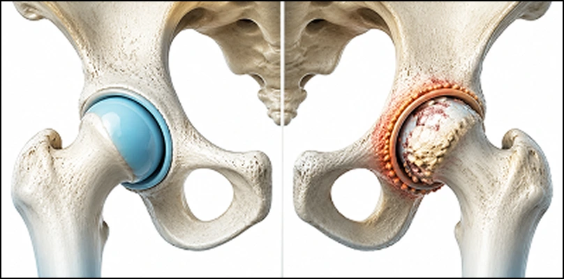
Hip osteoarthritis is called hip osteoarthritis or coxarthrosis in the medical literature. The hip joint is the surface between the femoral head (thigh bone) and the acetabulum (hip socket) in the pelvis. It is one of the body’s largest weight-bearing joints. In a healthy hip, the joint surfaces are covered with cartilage, which allows smooth motion.
In hip osteoarthritis, cartilage thins, wears, and may disappear. As cartilage is lost, the joint surfaces rub, and inflammation develops in the capsule and surrounding tissues. Pain, stiffness, limited motion, and loss of function follow. Early diagnosis and appropriate treatment are critical for preserving joint function and quality of life. As the disease advances, osteophytes (bone spurs) may form and the joint may deform.
Although often linked to aging, hip osteoarthritis can also appear at younger ages. Prof. Dr. Yavuz Arıkan emphasizes that congenital hip dysplasia, trauma, infection, and hip dislocation can accelerate osteoarthritis, so at-risk patients need regular follow-up. Hip osteoarthritis is therefore a public health issue that cannot be limited to “a disease of the elderly.”
What Causes Hip Osteoarthritis?
Many factors play a role. Cartilage degeneration usually starts when several factors combine, not from a single cause.
Aging and Cartilage Degeneration
With age, cartilage loses elasticity and repair capacity. The quality and amount of synovial fluid may also decline. Joint surfaces then wear faster.
Genetic Predisposition
People with a family history of osteoarthritis have a higher risk. Genetic factors can affect cartilage durability and joint stability.
Excess Weight and Overload
The hip is one of the body’s most load-bearing joints. Extra weight increases load and speeds cartilage wear. Prolonged standing, heavy lifting, and long walks can also increase cartilage damage.
Congenital or Early Hip Problems
- Congenital hip dislocation
- Hip dysplasia
- Perthes disease
- Slipped capital femoral epiphysis (epiphysiolysis)
These conditions disrupt normal development of the hip surfaces and set the stage for osteoarthritis.
Trauma and Joint Injury
Hip fractures, dislocations, and soft-tissue injuries can disrupt hip anatomy. Cartilage damage after trauma may later become osteoarthritis.
Inflammatory Diseases and Metabolic Disorders
Inflammatory joint diseases such as rheumatoid arthritis and psoriatic arthritis can cause chronic inflammation and cartilage loss. Metabolic diseases such as gout and hemochromatosis can also trigger osteoarthritis.
Symptoms of Hip Osteoarthritis
Symptoms usually develop slowly and at first appear only with activity. Patients often report:
- Pain in the groin or hip
- Pain that increases with weight-bearing
- Worsening pain after walking
- Difficulty going up and down stairs
- Stiffness in the morning or after sitting for a long time
- Limited hip motion (especially internal rotation and flexion)
- Limping or a gait change
- A “cracking” or “grinding” sound from the hip
Character of the Pain
Pain is most often felt in the groin. In some patients it radiates to the front of the thigh, the inner knee, or the side of the knee, so hip osteoarthritis can be mistaken for knee pain. According to Prof. Dr. Yavuz Arıkan’s clinical assessment in orthopedic oncology and bone disease, the pattern of radiation and a detailed history are important clues. Pain usually increases with movement and decreases with rest. In advanced stages it may continue at rest. Prof. Dr. Yavuz Arıkan emphasizes that early diagnosis and an appropriate plan are decisive for quality of life.
How Is Hip Osteoarthritis Diagnosed?
Diagnosis is based on history, physical examination, and imaging. The aim is to identify the source of pain and the stage of osteoarthritis.
History and Physical Examination
The physician asks about duration of symptoms, location of pain, aggravating and relieving factors, previous trauma, and medical history. Range of motion, tender points, and muscle strength are assessed. Tests such as the Trendelenburg test (hip abductor strength) and the FABER test (iliac/sacroiliac assessment) may be used.
Imaging
X-ray is the most common imaging method. Findings include:
- Joint-space narrowing
- Bone spurs (osteophytes)
- Subchondral sclerosis (bone hardening)
- Subchondral cysts
When needed, MRI can show cartilage, labrum, muscle, and soft tissue in more detail. Other causes of hip pain (for example femoroacetabular impingement or avascular necrosis) can also be excluded.
Stages and Progression of Hip Osteoarthritis
Hip osteoarthritis usually progresses slowly. Staging helps guide treatment. One of the most used systems is the Kellgren-Lawrence classification:
- Stage 1: Mild osteophytes, minimal joint-space narrowing
- Stage 2: Clear osteophytes, mild joint-space narrowing
- Stage 3: Moderate joint-space narrowing, subchondral sclerosis
- Stage 4: Severe joint-space loss, marked deformity, and cystic change
In early stages symptoms may be mild and daily activities can continue. In advanced stages pain and stiffness become marked, and gait disturbance and loss of function appear.
Treatment of Hip Osteoarthritis
The main goals are to reduce pain, preserve mobility, improve quality of life, and slow progression. Treatment should be individualized according to age, lifestyle, pain severity, and the degree of joint damage. Care usually combines conservative and, when needed, surgical approaches.
Conservative (Nonsurgical) Treatment
Weight Control and Lifestyle Changes
The hip carries a large share of body weight. Extra weight increases load and speeds cartilage wear. Weight control is one of the most effective approaches. Also helpful:
- Avoiding prolonged standing
- Not lifting heavy loads
- Walking on even, stable surfaces rather than soft ground
- Choosing appropriate shoes
Physical Therapy and Exercise
Strengthening the muscles around the hip improves stability and reduces pain. A physical therapy program may include:
- Hip abductor strengthening
- Hip flexor and extensor strengthening
- Core strengthening
- Stretching and mobilization
Swimming, water aerobics, and cycling are among the recommended low-impact activities.
Medication
The aim is short-term use at the lowest effective dose. Common options:
- Paracetamol (acetaminophen)
- NSAIDs (ibuprofen, naproxen, and similar)
- Topical pain relievers (creams, gels)
- Short courses of nonsteroidal anti-inflammatory drugs when needed
Stomach, kidney, and cardiovascular side effects must be considered, especially in older patients, so medical supervision is important.
Intra-articular Injections
Injections into the hip can reduce pain and inflammation. The most used methods:
- Corticosteroid injection (temporary pain control)
- Hyaluronic acid (joint lubricant) injection
- PRP (platelet-rich plasma) injection
These may help especially in early and moderate hip osteoarthritis. Effectiveness varies by patient and disease stage.
Surgical Options
Surgery is considered when conservative care cannot control pain and quality of life is seriously affected. Age, activity, disease stage, and general health are taken into account. Prof. Dr. Yavuz Arıkan emphasizes that correct patient selection and timing determine long-term success.
Arthroscopic Surgery
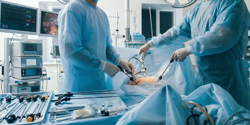
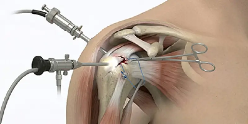
In early disease, if there is cartilage damage or femoroacetabular impingement, arthroscopy may be considered. Labrum repair, cartilage debridement, or corrective procedures can be done. In advanced osteoarthritis, arthroscopy is usually not enough.
Osteotomy (Bone Realignment)
In osteoarthritis related to structural problems such as hip dysplasia, osteotomy can improve load distribution and slow progression. It is usually preferred in younger and middle-aged patients.
Hip Replacement (Total Hip Arthroplasty)
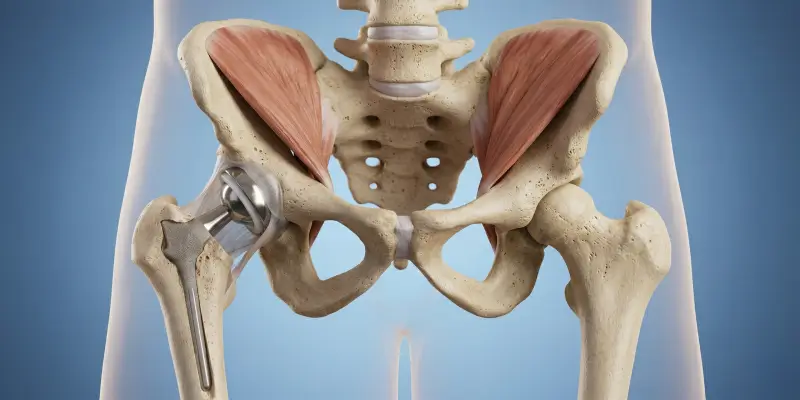
In advanced hip osteoarthritis, total hip replacement is the most effective and widely used treatment. The prosthesis replaces worn joint surfaces with an artificial joint. It substantially reduces pain and improves function.
Recovery varies with overall health. Walking usually begins within a few weeks, and return to daily activities often occurs in 6–12 weeks. In the long term, implant lifespan may be 15–20 years or longer.
Recovery and Rehabilitation After Treatment
Recovery depends on the method used and the patient’s general health. After conservative care, exercise and lifestyle changes continue. After surgery, rehabilitation is more structured.
Early Period After Surgery
- Pain control and early mobilization are the goals in the first days.
- Physical therapy starts gait training and muscle strengthening.
- Precautions are taken against falls and prosthesis dislocation.
Long-term Rehabilitation
- Strengthening muscles around the hip
- Walking and stair training
- Balance and proprioceptive exercises
- Return to daily activities
Regular exercise after hip replacement helps the implant last longer and preserves function.
What Happens If Hip Osteoarthritis Is Not Treated?
Untreated hip osteoarthritis progresses and seriously reduces quality of life. Over time:
- Pain becomes chronic
- Walking distance decreases
- Limping develops
- Hip motion becomes more limited
- Muscle atrophy (weakening) occurs
- Secondary problems may appear in the spine and knee
- In advanced stages, joint deformity and loss of function occur
Prolonged inactivity can also increase cardiovascular and metabolic risks. People with suspected hip osteoarthritis should be evaluated early.
Lifestyle: Which Sports and Exercises Are Safe?
Exercise is important, but the wrong activities can increase pain. Safer options include:
- Swimming and water exercise
- Low-impact cycling
- Walking (on even ground, starting with short distances)
- Balanced strength training (for hip and core muscles)
- Pilates (under physician/physiotherapist supervision)
Activities to avoid:
- Running and jumping (high impact)
- Heavy lifting
- Sudden turns and direction changes
- Prolonged squatting
Common Myths About Hip Osteoarthritis
Myth 1: “Osteoarthritis only happens in older people”
Fact: Hip osteoarthritis can also occur in younger people. Congenital hip dysplasia, trauma, and sports injuries can cause early osteoarthritis.
Myth 2: “Painkillers must be used continuously”
Fact: Long-term painkiller use can cause side effects. Priority should be weight control, exercise, and an appropriate treatment plan.
Myth 3: “A brace or support device corrects osteoarthritis”
Fact: Support devices may reduce pain, but they do not reverse osteoarthritis.
Conclusion
Hip osteoarthritis is a progressive, chronic joint disease that affects daily life. With early diagnosis, appropriate treatment, and lifestyle changes, pain can be controlled and function preserved. Weight control, regular exercise, muscle strengthening, and surgery when needed can substantially improve quality of life. Anyone with hip pain should be evaluated by a specialist and given an individualized plan.
Frequently Asked Questions

Specialist Opinion
Hip osteoarthritis is a progressive, chronic joint disease that affects daily life. With early diagnosis, appropriate treatment, and lifestyle changes, pain can be controlled and function preserved. Weight control, regular exercise, muscle strengthening, and surgery when needed can substantially improve quality of life. Anyone with hip pain should be evaluated by a specialist and given an individualized plan.
Contact us to have your symptoms assessed and to learn about treatment options that may be right for you. Get in touch .
Frequently Asked Questions
Nerves of the hip joint can cause pain that radiates into the thigh and knee. Some patients therefore feel hip-related pain in the knee.
Because osteoarthritis is a chronic disease, the goal is control rather than complete cure. In advanced stages, hip replacement can substantially improve pain and function.
A hip replacement often lasts 15–20 years or longer. Implant lifespan depends on the patient’s weight, activity level, and prosthesis type.
The decision is based on symptoms, quality of life, and disease stage. Age alone is not decisive; replacement can also be done in younger patients with advanced disease.
It cannot always be fully prevented, but reducing risk factors and early diagnosis can slow progression. Weight control, regular exercise, and avoiding hip trauma are important.
 WhatsApp
  Facebook
  Twitter
  Linkedin



 +90 505 398 34 34
 doctoryavuzarikan@gmail.com
 Book Appointment / Contact