

What Is Enchondroma? Symptoms, Diagnosis, and Treatment


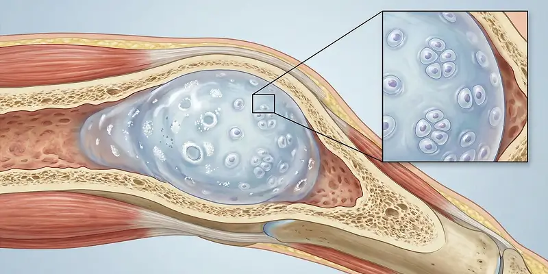
Enchondroma is a benign bone tumor caused by abnormal growth of cartilage cells. It is usually found in the middle and end portions of long bones, especially in the hands, feet, femur, and humerus. Enchondroma is most common in childhood and young adulthood, and many patients have no symptoms.
In some cases the tumor grows and weakens bone structure, causing microfractures, deformity, or chronic bone pain. Enchondromas in the fingers and toes can grow unnoticed for a long time and present with a pathologic fracture.
Most enchondromas are benign, but there is a rare risk of transformation into chondrosarcoma (a malignant cartilage tumor). Patients diagnosed with enchondroma should therefore be followed by an experienced orthopedic oncology team.
This article covers what enchondroma is, its symptoms, diagnostic methods, the risk of malignant transformation, and treatment.
What Is Enchondroma?
Enchondroma is a benign bone tumor that originates from cartilage and develops inside the marrow cavity. It is most common in the long bones of the hands and feet, especially the phalanges (finger bones), metacarpals, humerus (upper arm bone), and femur (thigh bone).
An enchondroma forms when cartilage cells remain inside bone during normal ossification and then multiply abnormally. The tumor sits as islands of cartilage within bone and can weaken the bone over time.
Most enchondromas grow slowly and can remain silent for years. Many patients therefore discover the tumor on an X-ray, MRI, or CT done for another reason. In some cases the tumor weakens bone, causes cracks or fractures, and produces pain.
Although most enchondromas are benign, large, painful, or rapidly growing lesions carry a risk of chondrosarcoma (cartilage-origin bone cancer). Every diagnosed enchondroma must therefore be kept under regular follow-up.
Enchondroma Symptoms
Enchondromas often cause no symptoms and can remain silent for a long time. When the tumor grows or the bone weakens, the following may appear:
- Bone pain: Deep bone pain felt especially during or after activity
- Swelling or a mass: Especially noticeable in the fingers and toes
- Pathologic fracture: Breakage after a light blow because the bone has weakened
- Limited motion: Loss of function when the enchondroma is near a joint
- Tenderness: Pain that increases with touch
Increasing pain, growing swelling, or night pain can be important warning signs of malignant transformation.
Diagnostic Methods
Diagnosis is based on clinical examination and imaging together. Biopsy is used when needed to confirm the diagnosis.
X-ray
The first diagnosis is usually made on X-ray. Well-defined, cartilage-origin lesions with calcification inside the bone are typical.
Magnetic Resonance Imaging (MRI)
MRI is used to assess how far the tumor has spread inside the bone, its relationship to soft tissues, and possible signs of malignant transformation.
Computed Tomography (CT)
CT more clearly shows thinning or destruction of the cortex. It is especially important for assessing fracture risk.
Biopsy
If the lesion is painful, growing rapidly, or looks suspicious, biopsy is used to examine cellular structure. This definitively distinguishes enchondroma from chondrosarcoma.
Differences Between Enchondroma and Chondrosarcoma
| Feature | Enchondroma | Chondrosarcoma |
|---|---|---|
| Cell structure | Benign cartilage | Malignant cartilage |
| Growth | Slow | Rapid and aggressive |
| Pain | Usually absent or mild | Severe pain that increases at night |
| Bone destruction | Limited | Spread into bone and soft tissue |
| Metastasis | None | Possible, especially to the lungs |
| Treatment | Observation or surgery | Wide surgery ± oncologic treatment |
In a sarcoma approach, if imaging shows aggressive findings, biopsy is used for a definitive distinction.
Enchondroma Fracture (Pathologic Fracture) Risk
Because enchondroma weakens the inside of the bone, even a very small blow can cause a pathologic fracture in:
- The fingers and toes
- The thigh bone
- The upper arm bone
At orthopedic oncology practices in Istanbul, the bone’s load-bearing capacity is calculated with specialized CT and MRI measurements.
When Should Enchondroma Be Operated On?
According to Prof. Dr. Yavuz Arıkan’s sarcoma protocols, surgery is recommended when:
- There is pain
- The tumor is growing
- The bone is weakened by more than 50%
- A fracture has occurred
- MRI shows aggressive signals
- Chondrosarcoma is suspected
At surgery the tumor is scraped out (curettage) and the cavity is filled with bone graft or a biologic filler.
Enchondroma Follow-up Protocol
At orthopedic oncology practices in Istanbul, enchondroma follow-up is standardized:
- First 2 years → MRI + X-ray every 6 months
- Later years → once a year
- Pain or growth → urgent re-evaluation
This system aims to catch transformation to chondrosarcoma early.
| Time | Check |
|---|---|
| 0–12 months | MRI + X-ray |
| 12–24 months | MRI |
| 2–5 years | Annual imaging |
| After that | Clinical follow-up |
Life After Enchondroma Surgery
After successful surgery:
- Walking returns to normal
- Hand and foot function is regained
- Sports and daily life are possible
- Patients who receive bone graft regain full strength within 3–6 months
Conclusion
Enchondroma is often benign, but it should never be taken lightly as a cartilage-origin bone lesion. Enchondromas in the hands, feet, femur, and humerus can remain silent for a long time, then present with pathologic fractures, deformity, and pain when the bone weakens.
At orthopedic oncology practices in Istanbul, and in Prof. Dr. Yavuz Arıkan’s practice, enchondromas are not simply labeled “benign.” They are followed closely for the risk of transformation to chondrosarcoma. According to the literature, some enchondromas can become malignant cartilage tumors, and this change usually first appears as increasing pain and radiologic changes.
The approach at centers experienced in sarcoma does not only assess the current tumor; it also covers future bone health, fracture risk, and possible malignant change. With regular imaging, well-timed surgery, and biopsy when needed, enchondromas can be controlled with a high success rate.
Most patients who are diagnosed early and enter an appropriate follow-up protocol can continue life without serious problems.
Frequently Asked Questions

Specialist Opinion
Enchondroma is often benign, but it should never be taken lightly as a cartilage-origin bone lesion. Enchondromas in the hands, feet, femur, and humerus can remain silent for a long time, then present with pathologic fractures, deformity, and pain when the bone weakens.
At orthopedic oncology practices in Istanbul, and in Prof. Dr. Yavuz Arıkan’s practice, enchondromas are not simply labeled “benign.” They are followed closely for the risk of transformation to chondrosarcoma. According to the literature, some enchondromas can become malignant cartilage tumors, and this change usually first appears as increasing pain and radiologic changes.
The approach at centers experienced in sarcoma does not only assess the current tumor; it also covers future bone health, fracture risk, and possible malignant change. With regular imaging, well-timed surgery, and biopsy when needed, enchondromas can be controlled with a high success rate.
Most patients who are diagnosed early and enter an appropriate follow-up protocol can continue life without serious problems.
Contact us to have your symptoms assessed and to learn about treatment options that may be right for you. Get in touch .
Frequently Asked Questions
No. Enchondroma is a benign cartilage tumor. Because some cases can transform into chondrosarcoma, it should be followed by sarcoma specialists.
Rarely, yes. Enchondromas that cause pain, grow rapidly, or disrupt bone structure can transform into chondrosarcoma. Orthopedic oncology specialists such as Prof. Dr. Yavuz Arıkan therefore recommend regular follow-up.
Most of the time it does not. Pain appears when the bone weakens, microfractures form, or the tumor grows.
Yes. Because enchondroma weakens bone, a pathologic fracture can occur even after minor trauma.
Not always. Small, painless lesions can be observed. Surgery is recommended for growing, painful enchondromas or those with fracture risk.
At surgery the tumor is curetted (cleared) and the cavity is filled with bone graft or a biologic filler. The procedure follows the principles of sarcoma surgery.
Usually for years. At orthopedic oncology practices in Istanbul, MRI and X-ray follow-up is more frequent in the first 2 years.
Rarely, yes. Incompletely cleared tumors can recur, which is why experienced orthopedic oncology surgery is important.
 WhatsApp
  Facebook
  Twitter
  Linkedin



 +90 505 398 34 34
 doctoryavuzarikan@gmail.com
 Book Appointment / Contact