

What Is an Aneurysmal Bone Cyst (ABC)? Symptoms, Diagnosis, and Treatment


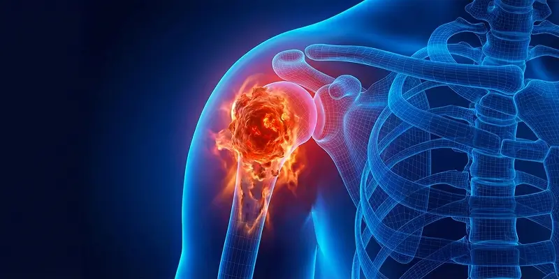
An aneurysmal bone cyst (ABC) is a bone lesion made of blood-filled spaces. It is considered benign but can grow aggressively. It is especially common in children and young adults and is clinically important because it weakens bone and increases fracture risk. ABC is not a true cancer, but it can disrupt bone structure and cause serious orthopedic problems.
This cyst type most often appears in people under 20, especially between ages 10 and 15. Among bone tumors in the pediatric and adolescent age group, aneurysmal bone cyst has an important place. Because it affects bone during rapid growth, it can have lasting effects on skeletal development.
Aneurysmal bone cysts most often involve large, load-bearing bones such as the femur (thigh bone), tibia (shinbone), pelvis, and spine. Lesions in these sites reduce mechanical strength and can cause progressive structural damage. Spinal ABCs can threaten both bone stability and the spinal cord and nerves.
ABC takes its name from enlarged, blood-filled vascular spaces inside the bone. These vessel-like cavities create pressure, erode normal bone, and replace it with cystic spaces. Over time the bone thins, expands, and weakens from within. On imaging this can look like a “blown-out” or ballooned bone.
Many aneurysmal bone cysts do not form an obvious mass at first and can grow unnoticed for a long time. As the lesion progresses, pressure and volume increase affect nearby bone, joints, and soft tissue. In load-bearing bones this seriously raises the risk of pathologic fracture.
The exact cause is not fully known. Factors thought to play a role include genetic predisposition, intraosseous circulation problems, microtrauma, and irregularities in bone metabolism. Some studies suggest ABC may develop when a bleed or hematoma inside bone organizes into a cystic structure.
In some cases, an aneurysmal bone cyst appears secondarily on top of another bone lesion. An underlying bone tumor can then trigger cyst formation and a more complex picture. ABCs often show both osteolytic (bone-destroying) and osteoblastic (new-bone-forming) activity, which reflects their dynamic, aggressive nature.
Aneurysmal Bone Cyst Symptoms
Clinical findings vary with the cyst’s size, the bone involved, growth rate, and the patient’s age. In early stages many patients are completely asymptomatic, and the lesion is found incidentally on an X-ray or MRI done for another reason. As the cyst grows and weakens bone, clear symptoms appear.
1. Pain
The most common symptom is bone pain. This pain usually:
- Starts insidiously and increases over time
- Worsens with physical activity
- May become prominent as night pain
The main cause is increased pressure inside the bone and erosion of healthy bone, which can lead to microcracks. ABCs in load-bearing bones such as the femur, tibia, and pelvis can cause severe pain.
2. Swelling and a Sense of a Mass
When the cyst expands the cortex, a visible or palpable swelling can form under the skin. This swelling:
- May feel firm or slightly elastic
- Can enlarge over time
- May be associated with increased warmth under the skin in some patients
This is more noticeable in superficial bones (tibia, humerus, bones of the hands and feet).
3. Limited Motion
If the ABC is near a joint, restricted joint motion can develop because of:
- Pressure on the joint capsule
- Reflex muscle spasm from pain
- Loss of bone stability
This can cause functional loss, especially in ABCs around the knee, hip, and shoulder.
4. Pathologic Fracture
Because aneurysmal bone cysts erode bone from within, strength is greatly reduced. A pathologic fracture can then occur after mild trauma or even during ordinary daily activity. These fractures are often the first reason the disease is noticed.
5. Neurologic Symptoms (Spinal Involvement)
When ABC involves the spine, more serious signs can appear:
- Numbness in the arms or legs
- Weakness
- Walking difficulty
- Rarely, problems with bladder or bowel control
These symptoms result from pressure on the spinal cord or nerve roots and require urgent care.
Diagnostic Methods
Reliable diagnosis is critical for planning treatment and distinguishing ABC from malignant bone tumors. Radiologically, ABC can mimic osteosarcoma, giant cell tumor, and chondrosarcoma. Diagnosis must therefore be led by an experienced orthopedic oncology team. The multidisciplinary approach used by Prof. Dr. Yavuz Arıkan at the orthopedic oncology practice in Istanbul supports accurate differential diagnosis.
The process usually includes:
1. Plain Radiograph (X-ray)
This is the first imaging step. On X-ray, an aneurysmal bone cyst typically shows:
- An expanded, ballooned area inside the bone
- A thin cortex
- A “soap-bubble” appearance
These findings are characteristic but not enough for a definitive diagnosis on their own.
2. Computed Tomography (CT)
CT more clearly shows:
- Damage to the cortex
- How much the bone has thinned
- Possible microfractures
It is also important for assessing bone integrity before surgical planning.
3. Magnetic Resonance Imaging (MRI)
MRI is the gold standard for diagnosing aneurysmal bone cyst because it clearly shows:
- Blood-fluid levels inside the cyst
- The fluid–fluid levels typical of ABC
- Soft-tissue extension
MRI helps determine whether the cyst is aggressive or limited and defines the extent of surgery.
4. Ultrasound
In ABCs of superficial bones, ultrasound can help assess fluid and blood flow inside the cyst. Its value is limited in deep bones.
5. Bone Scintigraphy
This is used to look for involvement of more than one bone or other active lesions. It is important when:
- Multifocal cysts are suspected
- Metastatic bone disease must be excluded
6. Biopsy (Definitive Diagnosis)
Aneurysmal bone cyst can look similar on imaging to malignant tumors such as chondrosarcoma, osteosarcoma, and giant cell tumor. Biopsy is therefore required for a definitive diagnosis.
During biopsy, a pathologist evaluates:
- Cyst contents
- Cellular structure
- Bleeding pattern
Oncologic biopsy techniques used by Prof. Dr. Yavuz Arıkan at the orthopedic oncology practice in Istanbul minimize the risk of misdiagnosis and spread.
Treatment Options
The plan depends on the cyst’s size, the bone involved, whether it is growing rapidly, pathologic fracture risk, and the patient’s age. The goal is not only to remove the cyst but also to preserve bone strength, reduce recurrence, and prevent loss of function. Orthopedic oncology protocols used by Prof. Dr. Yavuz Arıkan are based on this multifaceted approach.
1. Surgery (Complete Removal of the Cyst by Curettage)
The main treatment of ABC is surgical clearance (curettage). During this procedure:
- The cyst is opened into the bone
- All bloody and pathologic tissue is scraped out
- The cyst walls are cleaned with high-speed burrs
- Chemical or heat applications may be used to reduce recurrence
Surgery is the gold standard for ABCs that cause pain, are growing, or carry a fracture risk.
2. Arterial Embolization (Shrinking the Cyst by Cutting Off Blood Flow)
Arterial embolization blocks the vessels that feed the cyst. This method is preferred for:
- Very large ABCs
- High-risk sites such as the spine and pelvis
- Highly vascular cysts
Blood flow inside the cyst decreases, internal pressure drops, and the cyst shrinks over time. In some patients it can be definitive treatment; in others it is used to prepare for surgery.
3. Bone Grafting (Filling the Defect After Surgery)
The cavity left after curettage must be filled. Bone grafting is used here. The space may be filled with:
- The patient’s own bone (autograft)
- Donor bone (allograft)
- Synthetic bone-filling materials
This helps:
- Bone heal faster
- Reduce fracture risk
- Provide long-term strength
4. Radiofrequency Ablation
Radiofrequency ablation is a modern method used especially for small or hard-to-reach ABCs. A thin needle is placed into the cyst and:
- High heat is used to burn the cyst wall
- Bleeding is stopped
- Tumor cells are destroyed
This method is less painful than surgery and recovery is shorter.
5. Observation (Watch-and-Wait for Small, Asymptomatic Cysts)
Small ABCs that do not cause pain are not operated on immediately. In these patients:
- MRI or X-ray every 3–6 months
- Monitoring of pain and growth
is performed. If growth, pain, or bone weakening begins, active treatment is started.
Complications and Follow-up
Although an aneurysmal bone cyst is usually considered benign, late diagnosis or aggressive behavior can lead to serious complications. Long-term follow-up is therefore as important as treatment. In the orthopedic oncology approach used by Prof. Dr. Yavuz Arıkan, early recognition and prevention of complications are core goals.
1. Pathologic Fractures
ABC expands bone from within and thins the cortex. This can lead to a pathologic fracture:
- After mild trauma
- During daily activities
- Sometimes without trauma
The risk is higher in load-bearing bones such as the femur, tibia, and humerus. Pathologic fractures prolong treatment and increase the risk of functional loss.
2. Infection Risk
After surgery, though uncommon:
- Surgical-site infection
- Bone infection (osteomyelitis)
can develop. The risk is higher after extensive surgery and in recurrences that need reoperation. Infection signs should be watched closely in the early postoperative period.
3. Recurrence
One of the main problems with aneurysmal bone cysts is a tendency to recur. Recurrence is seen:
- Most often within the first 2 years
- Especially after curettage alone
Recurrence rates of 10–30% are reported in the literature. For that reason, Prof. Dr. Yavuz Arıkan prefers combined treatment methods in most patients.
4. Functional Losses
When ABC is near a joint or presents with pathologic fracture:
- Limited motion
- Loss of muscle strength
- Difficulty with daily activities
can occur. In children and young patients, involvement of growth plates can lead to long-term deformity.
How Should Follow-up Be Done?
Regular follow-up after treatment is essential for detecting complications and recurrence early. A typical protocol is:
- First 2 years: X-ray or MRI every 3–6 months
- Years 2–5: Imaging once a year
- If pain, swelling, or loss of function develops: urgent evaluation
During follow-up, activity level, pain, and bone strength are also assessed.
Conclusion
An aneurysmal bone cyst (ABC) is an important bone lesion seen especially in children and young adults. Although benign, it can weaken bone and cause serious functional problems. Early disease often has no symptoms, but later cases can produce pain, swelling, pathologic fractures, and limited motion that directly affect quality of life.
With modern methods, ABC can be distinguished from malignant bone tumors using X-ray, CT, MRI, and biopsy when needed. Because it can be confused with chondrosarcoma, osteosarcoma, and giant cell tumor, diagnosis at a center with orthopedic oncology experience is essential.
Treatment should be individualized with surgical curettage, bone grafting, arterial embolization, and radiofrequency ablation, according to the patient and the lesion. Multidisciplinary care at the orthopedic oncology practice in Istanbul, led by Prof. Dr. Yavuz Arıkan, aims both to reduce recurrence and to preserve bone function as much as possible.
The most important point is this:
Even though an aneurysmal bone cyst is benign, if left uncontrolled it can cause serious bone weakness, fractures, and lasting disability. Early diagnosis, appropriate treatment, and regular follow-up are the keys to safe management.
Frequently Asked Questions

Specialist Opinion
An aneurysmal bone cyst (ABC) is an important bone lesion seen especially in children and young adults. Although benign, it can weaken bone and cause serious functional problems. Early disease often has no symptoms, but later cases can produce pain, swelling, pathologic fractures, and limited motion that directly affect quality of life.
With modern methods, ABC can be distinguished from malignant bone tumors using X-ray, CT, MRI, and biopsy when needed. Because it can be confused with chondrosarcoma, osteosarcoma, and giant cell tumor, diagnosis at a center with orthopedic oncology experience is essential.
Treatment should be individualized with surgical curettage, bone grafting, arterial embolization, and radiofrequency ablation, according to the patient and the lesion. Multidisciplinary care at the orthopedic oncology practice in Istanbul, led by Prof. Dr. Yavuz Arıkan, aims both to reduce recurrence and to preserve bone function as much as possible.
The most important point is this: Even though an aneurysmal bone cyst is benign, if left uncontrolled it can cause serious bone weakness, fractures, and lasting disability. Early diagnosis, appropriate treatment, and regular follow-up are the keys to safe management.
Contact us to have your symptoms assessed and to learn about treatment options that may be right for you. Get in touch .
Frequently Asked Questions
No. An aneurysmal bone cyst is a benign bone lesion and is not cancer. Imaging findings can resemble some malignant bone tumors, so careful evaluation is required.
Some small, asymptomatic cysts may remain stable. Growing cysts that cause pain or weaken bone do not disappear on their own and need medical follow-up or treatment.
It is not directly life-threatening, but it can thin bone and lead to pathologic fractures, joint damage, and loss of function. Spinal cysts can compress nerves and cause serious neurologic problems.
Yes. There is a risk of recurrence, especially if the cyst is not fully cleared surgically. Regular MRI or X-ray checks after treatment are very important.
In some patients, minimally invasive methods such as arterial embolization or radiofrequency ablation can succeed without surgery. The right method depends on the cyst’s location and size.
Yes. They are most common in children and adolescents aged 10–20. Bone pain and swelling in childhood should therefore be taken seriously.
Yes. Because the cyst weakens bone, even simple trauma can cause a pathologic fracture. The risk is higher in the femur, tibia, and spine.
With appropriate treatment and follow-up, most patients can return to normal life and sports. Load-bearing should be limited for a time in high-risk areas.
Regular follow-up for at least 2–5 years after treatment is usually recommended. MRI or X-ray is used to check whether the cyst has returned.
Yes. Imaging can resemble osteosarcoma, giant cell tumor, or telangiectatic osteosarcoma. Evaluation at an experienced center is essential.
 WhatsApp
  Facebook
  Twitter
  Linkedin




 +90 505 398 34 34
 doctoryavuzarikan@gmail.com
 Book Appointment / Contact