

What Is a Simple Bone Cyst? Symptoms, Diagnosis, and Treatment


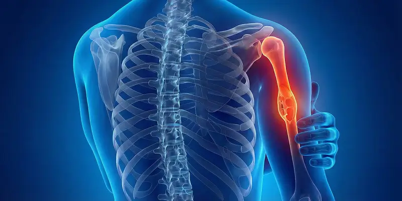
A simple bone cyst is a bone lesion caused by fluid collecting inside bone and is usually benign. It is most common in childhood and adolescence and tends to involve long bones such as the femur (thigh bone) and humerus (upper arm bone).
Simple bone cysts sit in the marrow cavity and are usually filled with clear or yellowish fluid. This fluid increases pressure inside the bone and weakens its internal structure. The bone becomes more fragile than normal, and a fracture can occur even without major trauma. This is called a “pathologic fracture.”
On X-ray, a simple bone cyst appears as a round or oval, well-defined empty area inside the bone. It is therefore often found incidentally on an X-ray done for another reason.
At orthopedic oncology practices in Istanbul, and in Prof. Dr. Yavuz Arıkan’s approach, simple bone cysts are evaluated not only as benign lesions but as bone weaknesses that carry a fracture risk. Untreated or growing cysts can lead to lasting bone deformity and repeated fractures in children and young adults.
Although simple bone cysts are not bone cancer, imaging findings can mimic some malignant bone tumors. Detailed imaging, biopsy when needed, and regular follow-up according to sarcoma principles are therefore important.
Causes of Simple Bone Cysts
The exact cause is not fully explained medically, but orthopedic oncology and sarcoma literature describe several factors that can trigger these lesions. At orthopedic oncology practices in Istanbul, and in Prof. Dr. Yavuz Arıkan’s clinical practice, simple bone cysts are especially linked to bone physiology during growth.
1. Genetic Predisposition
Familial predisposition is seen in some people. Those with a family history of bone cysts, bone weakness, or similar benign bone lesions have a higher chance of a simple bone cyst. This is related to genetic factors that affect fluid balance inside bone and bone formation.
2. Bone Development and the Growth Period
Simple bone cysts appear most often in childhood and adolescence. During this period bones lengthen rapidly and circulation in the marrow cavity changes. Increased fluid pressure inside bone can lead to cystic cavities. Sarcoma specialists describe this as a “fluid-drainage disorder related to the growth plate.”
3. Trauma and Micro-injuries
In some cases, cysts develop after blows to the bone, falls, or sports trauma. Trauma can cause small bleeds and fluid collection inside bone and set the stage for a cystic cavity. These cysts can later enlarge and weaken the bone.
4. Circulation Problems Inside Bone
One of the strongest theories is impaired venous drainage in the marrow cavity. Fluid collects inside bone, pressure rises, and bone tissue begins to resorb. This mechanism is assessed with advanced imaging at orthopedic oncology practices in Istanbul to determine fracture risk.
Symptoms
Simple bone cysts often cause no symptoms for a long time and are frequently found incidentally on an X-ray done for another reason. As the cyst grows, it weakens the internal structure of the bone and begins to press on nearby tissues. Various clinical signs can then appear.
At orthopedic oncology practices in Istanbul, and in Prof. Dr. Yavuz Arıkan’s practice, pain and fracture risk are assessed carefully because these findings indicate a need for active treatment.
1. Pain
When a simple bone cyst grows, pressure inside the bone increases and this can present as:
- Deep bone pain
- Pain that increases with exercise
- Sudden, severe pain if a fracture develops
Cysts in the femur and humerus can cause pain as load is applied.
2. Swelling and a Visible Mass
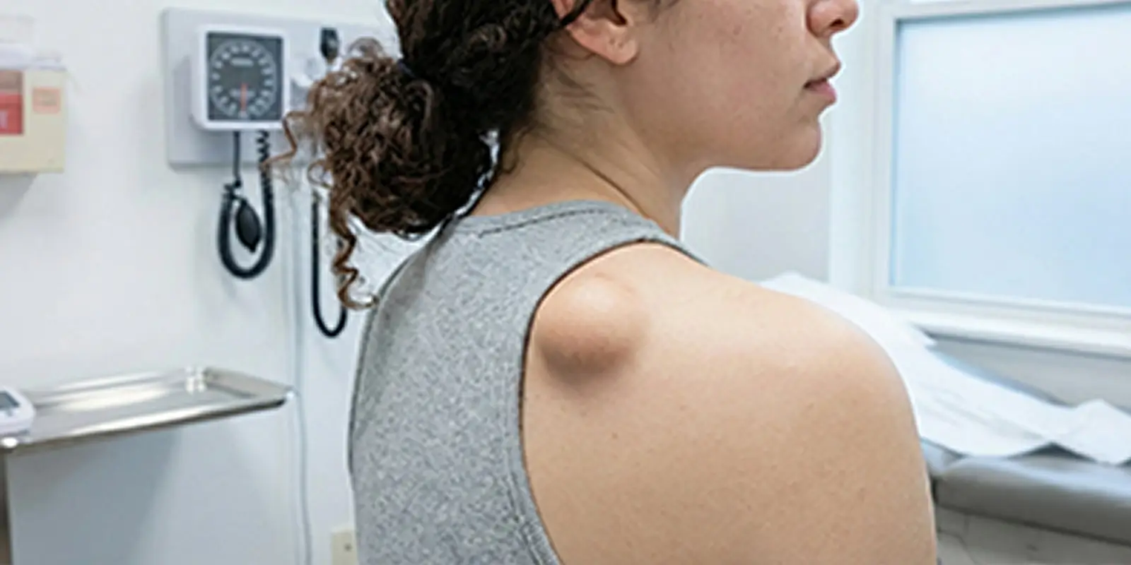
In bones close to the surface (arm, leg, around the shoulder), growth of the cyst can produce visible swelling or asymmetry. This can cause both cosmetic concern and loss of function.
3. Limited Motion
If the cyst is near a joint (knee, shoulder, hip), it can press on the joint capsule and muscles and cause:
- Reduced range of motion
- A feeling of stiffness
- Loss of strength
This can seriously affect sports in children and adolescents.
4. Pathologic Fracture
The most serious result of a simple bone cyst is pathologic fracture. Because the inside of the bone is emptied, a small blow, a fall, or sudden loading can cause a break. This is often the first noticed symptom.
Diagnostic Methods
Correct recognition of a simple bone cyst is essential for determining fracture risk and distinguishing it from malignant bone tumors. At orthopedic oncology practices in Istanbul, and in Prof. Dr. Yavuz Arıkan’s practice, diagnosis is not only about seeing the cyst; it includes analysis of bone strength and biologic behavior.
1. X-ray
This is the first-line imaging study. On X-ray the cyst appears:
- Well defined inside the bone
- Empty (radiolucent)
- Surrounded by a thin layer of bone
This typical appearance distinguishes a simple bone cyst from many other bone tumors.
2. Magnetic Resonance Imaging (MRI)
MRI clearly shows whether the contents are truly fluid and the relationship to nearby marrow, muscle, and joint tissue. It also answers:
- Is there bleeding inside the cyst?
- Is the wall thickened?
- Is there a tumoral structure?
This is vital from a sarcoma standpoint.
3. Computed Tomography (CT)
CT is used to determine how much the outer (cortical) bone has thinned and to assess fracture risk. It is indispensable for surgical planning, especially for cysts in the femur and humerus.
4. Biopsy (When Needed)
If imaging is suspicious or the cyst does not look typical, biopsy is used to examine cellular structure. This definitively distinguishes a simple bone cyst from an aneurysmal bone cyst or a malignant bone tumor.
Treatment
The goal is not only to shrink the cyst but to prevent fracture and stop recurrence. In approaches used by Prof. Dr. Yavuz Arıkan at the orthopedic oncology practice in Istanbul, treatment is planned according to the cyst’s location, whether the bone is load-bearing, and fracture risk.
1. Observation
Not every simple bone cyst needs treatment. Observation alone is enough when the cyst is:
- Small
- Painless
- Not weakening the bone
- Found incidentally
In these patients, X-ray or MRI at intervals is used to check:
- Is the cyst growing?
- Is the cortex thinning?
- Is a fracture risk developing?
In sarcoma follow-up protocols, growth plates in children are watched closely.
2. Aspiration and Injection
In this method:
- A needle is placed into the cyst
- The fluid is drained
- Healing substances are injected into the cyst
Possible injectates include:
- Corticosteroids
- Bone-stimulating substances
- Bone-marrow concentrate
The aim is to stop the cyst from producing fluid again and to start bone formation. This method is usually preferred:
- In children
- In cases where a solution without surgery is the goal
3. Surgery
Surgery is needed when:
- The cyst is large
- There is a risk of cracking or fracture
- A fracture has already occurred
- Aspiration has failed
What is done at surgery?
- The cyst is completely cleared (curettage)
- The cavity is filled with bone graft or synthetic bone material
- The bone is supported with metal implants if needed
Especially in load-bearing bones such as the femur and humerus, Prof. Dr. Yavuz Arıkan may perform fracture-prevention surgical stabilization.
Follow-up After Treatment
Simple bone cysts can also recur. Therefore:
- Close follow-up for the first 2 years
- Then annual imaging
is recommended. Sarcoma follow-up protocols monitor both cyst recurrence and bone strength.
Difference Between a Simple Bone Cyst and Bone Tumors
A simple bone cyst and bone tumors are often confused, but they are two different bone lesions with completely different biologic behavior, treatment needs, and risk. Correct differential diagnosis is essential to avoid unnecessary surgery and to catch dangerous tumors early.
What Is a Simple Bone Cyst?
A simple bone cyst is:
- A fluid-filled cavity
- Without cellular proliferation
- Not transforming into cancer
- A benign bone lesion seen mostly in children and young people
It usually forms during bone development and is not a tumor. It is often found incidentally.
What Is a Bone Tumor?
Bone tumors:
- Form from uncontrolled growth of bone cells
- May be benign (osteochondroma, osteoid osteoma, and similar lesions) or
- Malignant (chondrosarcoma, osteosarcoma, and similar tumors)
Malignant bone tumors destroy bone, can spread (metastasize), and can threaten life.
Main Differences
| Feature | Simple bone cyst | Bone tumor |
|---|---|---|
| Internal structure | Fluid-filled cavity | Cellular mass |
| Cancer risk | None | Possible |
| Growth pattern | Slow, passive | May be active and aggressive |
| Bone destruction | Minimal | May be marked |
| Spread | None | Present in malignant types |
| Treatment | Observation, aspiration, or surgery | Surgery ± oncologic treatment |
| Recurrence risk | Low to moderate | May be high depending on tumor type |
Why Is Differential Diagnosis So Important?
Simple bone cysts can sometimes resemble bone tumors on imaging. Especially in lesions that:
- Grow rapidly
- Cause pain
- Thin the cortex
- Cause fracture
the question “Is this really a cyst, or a tumor?” must be answered clearly.
In evaluations by Prof. Dr. Yavuz Arıkan at the orthopedic oncology practice in Istanbul, a definitive diagnosis is made with:
- MRI when needed
- CT
- Biopsy if required
Risks of a Wrong Diagnosis
If a bone tumor is mistaken for a “cyst”:
- Cancer is recognized late
- Treatment is delayed
- Limb loss or life-threatening results can occur
If a cyst is mistaken for a “tumor”:
- Unnecessary operations
- Psychological trauma
- Over-treatment
can occur.
Bone lesions should therefore be evaluated at centers specialized in musculoskeletal tumors, not with a routine orthopedic approach.
Conclusion
A simple bone cyst and bone tumors may look similar on imaging, but they are biologically completely different diseases. A simple bone cyst does not turn into cancer, while bone tumors can threaten life.
Every lesion found in bone should therefore be assessed by a specialist with the questions:
- “Can it be observed?”
- “Is surgery needed?”
- “Is there a cancer risk?”
The multidisciplinary approach used by Prof. Dr. Yavuz Arıkan at the orthopedic oncology practice in Istanbul protects patients from unnecessary treatment and helps catch possible cancers early.
Frequently Asked Questions

Specialist Opinion
A simple bone cyst and bone tumors may look similar on imaging, but they are biologically completely different diseases. A simple bone cyst does not turn into cancer, while bone tumors can threaten life.
Every lesion found in bone should therefore be assessed by a specialist with the questions:
“Can it be observed?” “Is surgery needed?” “Is there a cancer risk?”
The multidisciplinary approach used by Prof. Dr. Yavuz Arıkan at the orthopedic oncology practice in Istanbul protects patients from unnecessary treatment and helps catch possible cancers early.
Contact us to have your symptoms assessed and to learn about treatment options that may be right for you. Get in touch .
Frequently Asked Questions
No. Simple bone cysts do not turn into cancer. They are not tumors; they are benign cavities formed by fluid inside bone. Some bone tumors can look like cysts on imaging, so specialist evaluation is essential.
On its own it is usually not dangerous. If the cyst grows and weakens bone, it can lead to pathologic fractures. Close follow-up is needed especially in load-bearing bones (femur, humerus).
Most of the time it does not. Pain can appear if the cyst grows, the bone thins, or a fracture develops. Sudden pain is often a warning of fracture.
In some children, cysts may shrink or disappear as bone development is completed in adolescence. Not every cyst heals on its own; regular follow-up is essential.
Not always. Surgery is considered if the cyst is growing, the bone is weakened, there is a risk of repeated fracture, or there is pain. Prof. Dr. Yavuz Arıkan determines the need for surgery objectively with MRI and CT.
The cyst does not burst, but the bone containing the cyst can break. This causes pain and loss of function, and surgery may be needed.
X-ray, MRI, and CT are used to assess the internal structure, wall thickness, and bone destruction. Biopsy is done in suspicious cases.
Growth is usually monitored with X-ray every 6 months and MRI when needed.
If the cyst is small, usually yes. Large cysts carry a fracture risk, so impact sports such as football, basketball, and running may be restricted.
Yes. After aspiration or surgery it can recur in 10–30% of cases. Regular follow-up is therefore very important.
 WhatsApp
  Facebook
  Twitter
  Linkedin



 +90 505 398 34 34
 doctoryavuzarikan@gmail.com
 Book Appointment / Contact