

What Is a Glomus Tumor (Nail-Bed Tumor)? Symptoms and Treatment


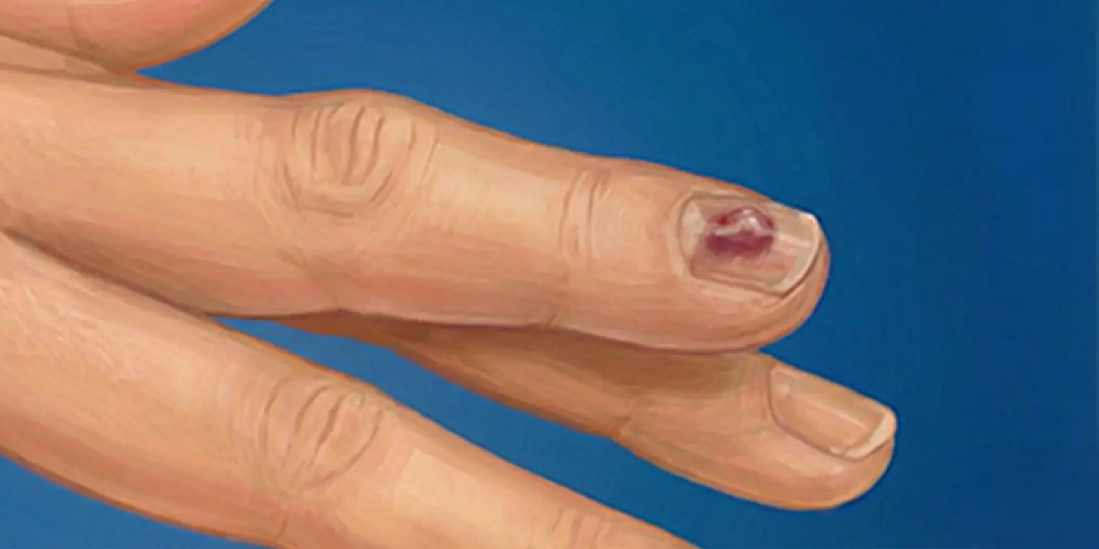
What Is a Glomus (Nail-Bed) Tumor?
A glomus tumor is a usually benign but extremely painful soft-tissue tumor that arises from glomus bodies in the nail bed and fingertips. Glomus bodies are specialized vascular structures that regulate heat balance and blood flow, especially in the fingers and toes. A glomus tumor that develops from these structures causes severe pain despite its small size because of its dense nerve and vessel content.
The nail bed is rich in nerves and vessels, so glomus tumors are most common there. They may be only millimeters in size, but the pain can seriously affect daily life. Stabbing, throbbing pain that increases with cold is the most characteristic feature. Many patients therefore receive incorrect treatments for a long time, thinking they have cold sensitivity, an ingrown nail, or rheumatic pain.
On imaging, a glomus tumor appears as a small mass under the nail with dense vascular structure. When it sits very close to bone, it can in some cases be confused with a bone tumor or a bone cyst. Correct diagnosis therefore requires evaluation from the perspective of hand surgery and orthopedic oncology.
In the orthopedic oncology approach advocated by Prof. Dr. Yavuz Arıkan, early detection of these small but high-impact tumors under the nail bed with the right imaging avoids unnecessary biopsies and incorrect bone procedures. With early diagnosis, glomus tumors can be completely removed with a simple operation, and the patient’s pain can be ended permanently.
Why Does a Glomus Tumor Form?
The exact mechanism is not fully known, but the structural and vascular features of glomus bodies in the nail bed and fingertips play a central role. Glomus bodies are specialized microvascular structures that regulate blood flow in the body’s extremities and respond to temperature change. Uncontrolled growth or abnormal vascular expansion of these structures can over time lead to a glomus tumor.
One of the most important causes is microtrauma. The nail bed is constantly exposed to door strikes, crushing, blows, manicure procedures, or repeated pressure. These small injuries can damage glomus bodies and cause abnormal cell growth during healing. Over time this process can become a benign but painful tumor.
Another important factor is cold exposure. Because glomus bodies are involved in heat regulation, they become more active in cold environments. Long-term cold exposure can overstimulate these cells and contribute to vascular expansion and tumor formation. Marked cold sensitivity in patients with a glomus tumor is therefore not a coincidence.
In rare cases genetic predisposition may play a role. In familial glomus tumor cases, multiple lesions can develop in more than one finger or in different parts of the body. In these patients the tumors usually appear at a younger age.
In some cases a glomus tumor sits so close to bone on imaging that it can be confused with a bone tumor. Diagnosis should therefore evaluate not only the presence of a mass but also the tissue of origin.
Where Do Glomus Tumors Occur?
Glomus tumors are most common in the nail beds of the fingers and toes. The main reason is that glomus bodies are densest in the fingertips and under the nails. These areas must respond quickly to temperature change and therefore have a rich network of vessels and nerves. This dense structure makes it easier for glomus tumors to develop here.
The most common sites are:
Under the fingernails (subungual region): This is the most frequent site. A small mass under the nail presents with severe pain and cold sensitivity. Because glomus tumors here sit very close to bone on X-ray and MRI, they can sometimes raise suspicion of a bone tumor.
Toes: Especially the big toe and second toe. Pain can become more noticeable because of increased pressure while walking.
Fingertips and soft tissues: Besides the nail bed, they can develop on the sides of the fingertip or in the soft tissues. These glomus tumors are sometimes confused with nerve or vessel tumors.
Palm and sole: Less common, but tumors from glomus bodies in these areas can cause severe pain that increases with pressure.
Rare sites: Very rarely, glomus tumors have been reported in internal organs such as the ear, stomach, and lung. The nail bed and fingertips remain the most typical sites.
Location directly affects the severity of symptoms and the diagnostic process. Tumors under the nail bed sit so close to bone that they can be mixed with bone-origin pain, which requires a detailed orthopedic oncology evaluation.
Glomus Tumor Symptoms
Although a glomus tumor is small, it is one of the most painful benign tumors in the body. The reason is its dense nerve endings and vascular structure. When it sits under the nail bed, distinctive, characteristic symptoms appear that seriously affect daily life.
The three most typical symptoms are:
1. Severe, stabbing pain: The pain is usually described as a pinprick, burning, or electric shock. It comes in short but very intense attacks. A simple touch, the tip of a pen, or light pressure on the nail can trigger it.
2. Extreme sensitivity to cold: Cold air, cold water, or contact with a metal surface suddenly increases the pain. Patients often feel the need to keep the hand away from cold. This feature is an important clue that distinguishes a glomus tumor from many other diseases.
3. Pinpoint tenderness: The patient can clearly show that the pain comes from a single point. Pressing a specific area of the nail bed dramatically increases the pain.
Other symptoms: Blue-purple or reddish color change under the nail, nail deformity over time, a local feeling of swelling or fullness, constant discomfort, and interrupted sleep.
In some patients the pain remains unexplained for a long time and can be confused with bone-origin problems or a bone tumor. This confusion is more common with glomus tumors under the nail bed because they sit so close to bone. Correct evaluation should therefore look at all of these distinctive signs together, not only at pain.
Difference Between a Glomus Tumor and a Bone Tumor
A glomus tumor and a bone tumor can resemble each other in both symptoms and imaging when they appear in the nail bed and fingertips. The two diseases, however, are completely different in origin, behavior, and treatment.
1. Tissue of Origin
A glomus tumor develops from glomus bodies made of vascular and nerve-rich structures. It is therefore of soft-tissue and vessel origin.
A bone tumor arises directly from bone tissue, bone marrow, or cartilage cells. This difference determines how the tumor grows and how it affects the body.
2. Size and Behavior
Glomus tumors are usually a few millimeters in size; despite being small they cause very severe pain and grow by pushing nearby tissues aside.
Bone tumors can form larger masses, disrupt the internal structure of bone, and over time weaken the bone or cause deformity.
3. Pain Features
In a glomus tumor the pain is sudden, sharp, and pinpoint; it increases clearly with cold and intensifies with pressure.
In a bone tumor the pain is deeper and duller; it usually increases at night and becomes constant over time.
4. Imaging Differences
On MRI and X-ray, a glomus tumor appears as a small but densely vascular mass under the nail bed. A bone tumor appears as a lesion that destroys, erodes, or hardens bone structure. When a glomus tumor sits very close to bone, it can be perceived as a bone-origin problem.
5. Treatment Approach
A glomus tumor heals permanently when it is completely removed with a simple but delicate operation. A bone tumor, depending on type, may require wide surgery, bone reconstruction, and sometimes chemotherapy or radiotherapy.
How Is a Glomus Tumor Diagnosed?
Diagnosis is possible because of very distinctive symptoms despite the small size; if these signs are not interpreted correctly, patients can be treated with the wrong diagnosis for a long time. Glomus tumors under the nail bed can be mixed with bone pain, nerve entrapment, or even suspicion of a bone tumor, so the diagnostic process must be careful and systematic.
1. Clinical Evaluation
The first step is a detailed history. The physician specifically asks about pain that increases with cold, pinpoint tenderness, and sudden, severe pain attacks. When this triad is present together, a glomus tumor is strongly considered. On examination it is typical for the patient to point exactly to the pain when a specific point on the nail bed is pressed.
2. Imaging Methods
Magnetic resonance imaging (MRI): This is the gold standard for diagnosing a glomus tumor. On MRI it appears as a small, well-defined, vascular mass under the nail bed. Proximity to bone and whether there is bone damage are also assessed.
Ultrasound: Can be used to show vascular structure and the mass in superficial glomus tumors.
X-ray: Often normal. If the tumor has pressed on bone for a long time, a superficial depression in the bone may be seen. This finding can sometimes be misinterpreted as a bone tumor.
3. Differential Diagnosis
A glomus tumor can be confused with: an ingrown nail, nerve tumors, soft-tissue cysts, and bone tumors. Evaluation should therefore look not only at pain but at imaging findings and the tumor’s vascular structure together. An orthopedic oncology approach prevents misdiagnosis at this point.
Treatment of a Glomus Tumor
The main and most effective treatment is complete surgical removal of the tumor. Although these tumors are benign, their dense nerve and vessel content causes very severe pain, so leaving them untreated seriously reduces quality of life.
1. Surgery
The gold-standard treatment is complete removal of the tumor with a delicate operation from the nail bed or the site involved. The aims of surgery are to clear all tumor tissue, protect healthy nerves and vessels, and preserve the natural structure of the nail bed as much as possible.
For glomus tumors under the nail, the surgeon usually reaches the mass by partially lifting the nail or approaching from the side. When the tumor is removed together with its capsule, the pain usually disappears completely immediately after surgery.
2. Incomplete Removal and Recurrence Risk
The most important risk with glomus tumors is that part of the tumor remains inside. If the mass is not fully removed, pain can continue and the tumor can grow again over time. Surgery should therefore be done by experienced hands and with appropriate imaging.
3. After Surgery
Most patients are discharged the same day or the next day, return to daily life in a short time, and the nail bed returns toward normal within a few weeks.
4. Are Radiotherapy or Drugs Needed?
Because glomus tumors are benign, chemotherapy, radiotherapy, or long-term drug treatment is not required. When completely removed by surgery, they are treated permanently.
Conclusion
A glomus (nail-bed) tumor is often very small, yet it is one of the benign tumors that most reduce quality of life. These tumors, which originate from glomus bodies under the nail and in the fingertips, cause severe, stabbing pain that increases with cold because of their dense nerve and vessel content. If these distinctive signs are not interpreted correctly, patients can receive incorrect treatments for a long time for an ingrown nail, rheumatism, nerve entrapment, or even a bone tumor.
The location of a glomus tumor directly affects both the severity of symptoms and the diagnostic process. Lesions that develop under the nail bed very close to bone can be perceived on imaging as a bone-origin problem. Correct diagnosis therefore requires detailed clinical evaluation, advanced imaging such as MRI, and interpretation from an orthopedic oncology perspective together. This approach prevents unnecessary biopsy and major bone surgery.
From a treatment standpoint, a glomus tumor is a disease that heals permanently when completely removed by surgery. Removing the tumor completely together with its capsule both makes the pain disappear immediately and eliminates the risk of recurrence. After surgery with the appropriate technique, patients can return to daily life in a short time, and nail structure is largely preserved.
In summary, when a glomus tumor is recognized correctly and treated with the right method, the results are highly rewarding. The most important point is to notice this small but high-impact tumor early, without confusing it with a bone tumor or other diseases, and to treat it in a targeted way. In this way patients can be freed from years of unnecessary pain and uncertainty and return to a healthy life.
Frequently Asked Questions
Frequently Asked Questions
A glomus tumor is a usually benign but very painful soft-tissue tumor that develops from glomus bodies in the nail bed and fingertips.
No. The large majority of glomus tumors are benign. Malignant glomus tumors are extremely rare.
Because it contains dense nerve endings and vascular structures. Even a light touch, pressure, or contact with cold can cause severe pain.
It is most common under the fingernails. It can also appear in the toes, fingertips, and, rarely, the palm and sole.
No. A glomus tumor is vascular and soft-tissue in origin. When it sits very close to bone, it can be confused with a bone tumor.
Diagnosis is based on clinical signs (cold sensitivity, pinpoint pain) and magnetic resonance imaging (MRI). MRI clearly shows the tumor’s location and vascular structure.
No. It does not disappear without treatment, and the pain usually increases over time.
The definitive treatment is surgery. When the tumor is completely removed, the pain disappears permanently.
If the tumor is completely removed, the recurrence risk is very low. If removal is incomplete, it can come back.
It usually does not pose a risk to life, but severe pain can seriously reduce quality of life. With correct diagnosis and treatment it can be fully cured.
 WhatsApp
  Facebook
  Twitter
  Linkedin




 +90 505 398 34 34
 doctoryavuzarikan@gmail.com
 Book Appointment / Contact