

What Is Synovial Chondromatosis? Symptoms, Diagnosis, and Treatment


What Is Synovial Chondromatosis?
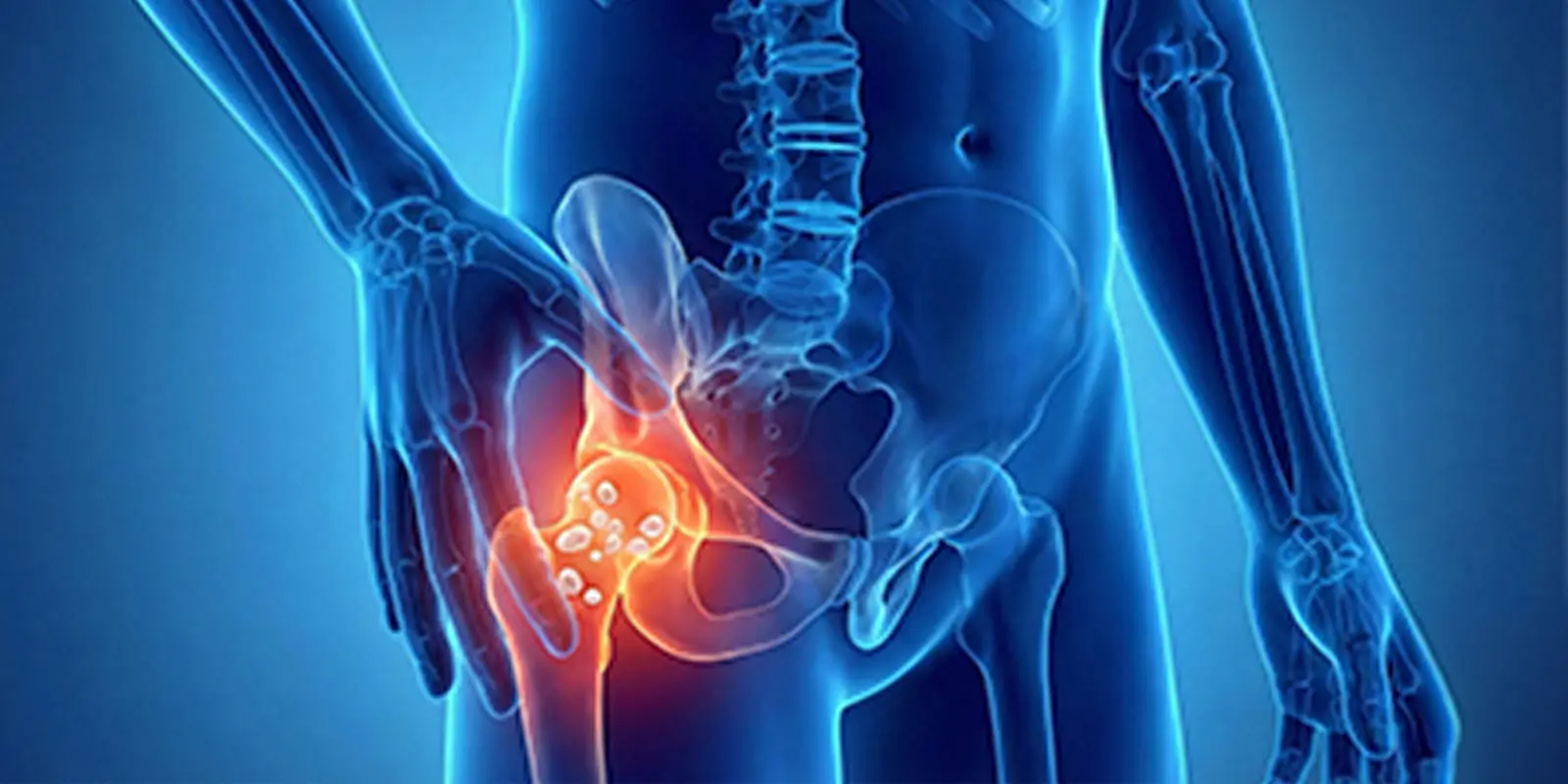
Synovial chondromatosis is a rare joint disease that appears when the synovial lining covering the inner surface of joints begins to produce cartilage abnormally. Small islands of cartilage form inside the synovial lining, and over time these structures break off into the joint space. Once free inside the joint, these cartilage fragments cause friction, catching, and pain during movement.
The disease is technically considered benign, but its behavior can be quite destructive. The hard cartilage fragments circulating inside the joint wear down healthy joint surfaces and over time can lead to arthritis (osteoarthritis). When it develops in the knee, hip, shoulder, and elbow, walking, raising the arm, or daily movements can be markedly restricted.
Synovial chondromatosis often progresses slowly. At first there may be only mild fullness and occasional pain; later, joint locking, catching during movement, sudden pain attacks, and swelling with stiffness appear. This picture can in some patients be confused with rheumatism, a meniscus tear, or even a bone tumor.
On imaging, many round cartilage or calcified nodules inside the joint are typical of synovial chondromatosis. MRI and CT show that these masses originate from the synovial lining, not from bone. This distinction is extremely important in orthopedic oncology for correctly separating intra-articular tumors from bone tumors.
In summary, synovial chondromatosis is not a “simple joint disease”; if untreated it can lead to lasting joint damage and advanced loss of motion. When it is noticed early and handled correctly, the joint can be preserved and patients can return to normal life.
Why Does Synovial Chondromatosis Form?
Synovial chondromatosis appears when the synovial lining covering the inner surface of the joint leaves its normal role and begins to produce cartilage abnormally. Normally this lining only produces joint fluid and allows the joint to move smoothly. In this disease, synovial cells begin to form cartilage-like tissues in an uncontrolled way. These cartilage foci grow, harden, and break off into the joint as loose bodies.
The exact trigger cannot always be identified, but important factors known to play a role in synovial chondromatosis include the following.
1. Metaplasia of Synovial Cells
The basic mechanism is that synovial lining cells undergo a structural change and turn into cartilage cells. This biologic change is called “metaplasia.” Once it starts, the joint begins to produce cartilage inside itself, and the process does not stop on its own.
2. Repeated Joint Trauma
Especially in frequently used joints such as the knee, shoulder, and elbow, sports injuries, falls, heavy loading, and occupational strain cause constant irritation of the synovial lining. This chronic irritation can change cell structure and trigger synovial chondromatosis.
3. Inflammation and Degeneration Inside the Joint
In some patients, osteoarthritis (arthritis), rheumatoid arthritis, and inflammation inside the joint disrupt the structure of the synovial lining and set the stage for this disease. This is called “secondary synovial chondromatosis.”
4. Impaired Blood Flow and Tissue Nutrition
The synovial lining needs regular, balanced blood flow to stay healthy. Impaired blood supply means the cells cannot get enough oxygen and nutrients. In such an environment, the normal biologic control mechanisms of synovial cells weaken, and the cells may produce cartilage-like tissue instead of their proper structure. Over time this abnormal production sets the stage for cartilage nodules accumulating inside the joint and for synovial chondromatosis.
Which Joints Does Synovial Chondromatosis Affect?
Synovial chondromatosis can theoretically develop in any joint that contains a synovial lining; clinically, however, some joints are affected much more often. The main reason is that these joints both carry a high movement load and have synovial linings with a larger surface area.
Most Commonly Affected Joints
Knee joint: This is the joint in which synovial chondromatosis is most common. The knee is the body’s most load-bearing joint and has the largest synovial space. Cartilage fragments inside the joint cause catching, locking, swelling, and pain while walking. On imaging they can sometimes resemble a bone-origin mass and be confused with a bone tumor.
Hip joint: Because it sits deep, diagnosis can be delayed. When synovial chondromatosis develops in the hip, groin pain, limp, and limited motion come to the fore. If it is not noticed early, lasting cartilage damage can develop in the hip joint.
Shoulder joint: Loose cartilage bodies in the shoulder cause catching and sudden pain when raising the arm. In some patients it can be confused with frozen shoulder.
Elbow joint: Cartilage fragments accumulating inside the elbow cause stiffness and locking during opening and closing. In advanced cases, wear of the bone surfaces is seen.
Why Are These Joints Affected More?
In frequently used joints that work under high load, the synovial lining is irritated more, microtrauma is more common, and circulation inside the joint is more variable. These factors make it easier for the synovial lining to start producing cartilage.
Less Commonly Affected Joints
Synovial chondromatosis can more rarely appear in small joints such as the wrist, ankle, jaw joint, and finger joints.
Symptoms of Synovial Chondromatosis
Synovial chondromatosis results from abnormal cartilage production by the synovial lining inside the joint and can over time seriously disrupt joint function. Symptoms are therefore not only simple joint pain; as the disease progresses it can create a complex picture that may be confused with a bone tumor, arthritis, and even joint locking.
1. Joint Pain (The Earliest and Most Common Symptom)
The first symptom of synovial chondromatosis is usually deep, dull joint pain. This pain increases with movement, intensifies after standing for a long time, and can reach a level that wakes the patient at night. Pain in the knee, hip, and shoulder in particular leads many patients to see a doctor with suspicion of a bone tumor.
2. Catching and a Feeling of Locking Inside the Joint
As synovial chondromatosis progresses, cartilage fragments inside the joint become free. These loose bodies catch during joint movement, cause sudden locking, and create a “caught” feeling while walking or raising the arm. This is especially noticeable in knee and shoulder involvement.
3. Swelling and Shape Change of the Joint
Cartilage nodules accumulating in the joint space and thickening of the synovial lining can cause swelling that is visible from the outside. This swelling increases over time, is sometimes felt as a firm mass, and can create an appearance that may be confused with a bone tumor. It becomes especially noticeable in the knee and ankle.
4. Limited Motion
Loose cartilage fragments inside the joint and synovial thickening narrow the joint space, damage cartilage surfaces, and over time lead to lasting stiffness. The patient cannot fully straighten the knee, bend the hip comfortably, or raise the shoulder.
5. Sounds From the Joint (Crepitus)
Sounds such as clicking, rubbing, and ticking may be heard during movement. These sounds come from loose cartilage fragments rubbing between the joint surfaces.
6. Joint Swelling Without Inflammation
Synovial chondromatosis most often causes swelling without redness, without fever, and without signs of infection. This is important in distinguishing it from rheumatic diseases.
How Is Synovial Chondromatosis Diagnosed?
Synovial chondromatosis cannot be diagnosed from the patient’s complaints alone. The disease can produce symptoms similar to a bone tumor, arthritis, ligament injuries, and loose bodies inside the joint. Correct diagnosis therefore requires clinical examination and advanced imaging together.
1. Clinical Examination and History
Diagnosis begins with a detailed history. The doctor specifically asks about the duration of joint pain, a feeling of catching or locking, whether swelling is increasing, and limited motion. Physical examination assesses firmness inside the joint, sounds during movement, and swelling. At this stage the disease can create a picture that raises suspicion of a bone tumor.
2. X-ray (Plain Radiograph)
X-ray is the first imaging method used in diagnosing synovial chondromatosis. In advanced disease, many small calcified nodules inside the joint, a “pyramid” or “grainy” appearance, and irregularity of the joint space may be found. In the early period, however, cartilage nodules may not yet be calcified, and the X-ray can look normal.
3. Magnetic Resonance Imaging (MRI)
MRI is the gold standard for diagnosing synovial chondromatosis. MRI shows non-calcified cartilage nodules, thickening of the synovial lining, and the number and location of loose bodies inside the joint in detail. Whether the disease has damaged bone tissue is also assessed. This allows differential diagnosis from a bone tumor.
4. Computed Tomography (CT)
CT clearly shows calcified cartilage nodules and wear of the bone surface. It is often used before surgical planning.
5. Differential Diagnosis (Can Be Confused With a Bone Tumor)
On imaging, synovial chondromatosis can sometimes appear as masses extending into bone. It can then be confused with osteochondroma, chondrosarcoma, and other bone tumors. Experienced orthopedic oncology evaluation is therefore very important.
6. Is Biopsy Needed?
In most cases MRI and CT findings are enough for diagnosis. If imaging results are suspicious or a malignant bone tumor cannot be excluded, a controlled biopsy can be done. Biopsy clarifies the diagnosis especially in atypical or aggressive-looking lesions.
Treatment of Synovial Chondromatosis
Synovial chondromatosis does not resolve on its own and over time can cause lasting damage inside the joint. Treatment is therefore planned not only to reduce pain but to stop abnormal cartilage formation inside the joint and to protect the joint. The right approach depends on the stage of disease, the joint involved, and the number of cartilage fragments.
1. Is Treatment Needed?
Synovial chondromatosis is usually a progressive disease. Cartilage nodules inside the joint grow over time, become free, damage the joint cartilage, and lead to arthritis. Lasting recovery is therefore usually not achieved in patients followed with medication alone. If untreated, the disease can result in bone damage and serious loss of motion in later stages.
2. Medication
Drugs do not eliminate synovial chondromatosis, but they reduce pain, control inflammation, and provide relief before surgery. Painkillers, anti-inflammatory drugs, and injections into the joint are used. These treatments do not destroy the cartilage nodules; they only suppress symptoms.
3. Surgery (The Main Treatment)
The definitive treatment of synovial chondromatosis is surgery. Surgery has two main aims:
- Removal of all loose cartilage fragments inside the joint
- Clearance of the synovial lining that produces abnormal cartilage (synovectomy)
Arthroscopic (minimally invasive) surgery: Done through small incisions; a camera is used to enter the joint, loose cartilage fragments are cleared, and the synovial lining is partly removed. It is especially effective in the knee and shoulder. Recovery time is short.
Open surgery: Open surgery is needed if the nodules are very large, extend behind the joint, or have worn the bone surface. In this method the joint is opened directly and all diseased tissue is cleared.
4. Recurrence Risk
One of the most important problems in synovial chondromatosis is the risk of recurrence. If the synovial lining is not cleared enough, the disease can start again and new cartilage nodules can form. During surgery it is therefore very important to remove not only the loose fragments but also the diseased synovial lining.
5. Rehabilitation After Surgery
After surgery, physical therapy, range-of-motion exercises, and muscle strengthening are used. The aim is to protect the joint and prevent stiffness.
Conclusion
Synovial chondromatosis is a disease that appears when the synovial lining inside the joint produces cartilage abnormally. It progresses insidiously and, if untreated, can lead to serious joint damage. At first it may present only with mild pain and a catching feeling; over time, accumulation of loose cartilage bodies inside the joint can lead to locking, swelling, limited motion, and lasting cartilage destruction. This picture both makes daily life harder and can cause the disease to be confused with more serious problems such as a bone tumor.
In diagnosis, advanced imaging methods, especially MRI, are very important; X-rays can look normal in the early period, while growing cartilage nodules inside the joint can be found only with detailed studies. Correct diagnosis prevents unnecessary treatments and allows timely surgery to protect the joint.
The essence of treatment is not only to reduce pain but to clear all loose cartilage fragments inside the joint and the diseased synovial lining. With appropriate surgery and physical therapy afterward, many patients can return to a life without pain and with preserved motion. Because the disease can recur, however, regular follow-up and early intervention are extremely important.
In summary, synovial chondromatosis can permanently damage the joint if neglected, but it can be fully controlled with early diagnosis and the right treatment. Anyone with long-lasting joint pain, catching, and swelling should not ignore this possibility; that is the most important step in protecting the joints.
Frequently Asked Questions
Frequently Asked Questions
Synovial chondromatosis is a disease in which the synovial lining that surrounds a joint produces cartilage abnormally, forming many small cartilage fragments inside the joint. These fragments harden over time and can disrupt joint motion.
No. Synovial chondromatosis is not a bone tumor. On imaging it can appear to extend into bone, so it can be confused with some bone tumors. Correct diagnosis is therefore very important.
It is most common in the knee. The hip, shoulder, and elbow are also frequently affected. More rarely it can appear in small joints such as the wrist and ankle.
The exact cause is unknown. Structural disorders of the synovial lining, irritation inside the joint, trauma, and impaired tissue nutrition may play a role.
No. The disease is usually progressive. Cartilage fragments inside the joint increase over time and damage the joint cartilage. Without treatment, lasting joint damage can occur.
The most important diagnostic method is MRI. MRI shows non-calcified cartilage nodules, thickening of the synovial lining, and loose bodies inside the joint in detail. X-ray and CT can also help.
Drugs can reduce pain but do not eliminate the disease. For lasting treatment, most patients need surgery to remove the cartilage fragments inside the joint and the diseased synovial lining.
Yes, synovial chondromatosis can recur. Adequate clearance of the synovial lining during surgery and regular follow-up afterward are therefore very important.
If untreated, it damages the joint cartilage and can cause serious arthritis at a young age.
Yes. With appropriate surgery and regular physical therapy, most patients can walk without pain, use their joints normally, and return to daily life.
 WhatsApp
  Facebook
  Twitter
  Linkedin




 +90 505 398 34 34
 doctoryavuzarikan@gmail.com
 Book Appointment / Contact