

What Is Lumbar Disc Herniation? Symptoms and Treatment


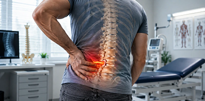
What Is Lumbar Disc Herniation?
Lumbar disc herniation occurs when the outer layer of the disc between the vertebrae (annulus fibrosus) tears and the gel-like inner core (nucleus pulposus) bulges outward. This bulge can compress nearby nerve roots and cause pain, numbness, tingling, and weakness. Disruption of disc structure can also create imbalance in the spinal segment and lead to chronic pain. Discs act as cushions connecting the mobile segments of the spine. Their roles are shock absorption, flexibility, and load distribution. When disc structure is damaged, imbalance and pain appear.
Lumbar disc herniation is most common at L4–L5 and L5–S1, because these regions move the most and carry the most load. It is more common between ages 30 and 50, but it can also occur in younger and older people. Lumbar disc herniation is not only pain; it is a serious health problem that can reduce quality of life, cause loss of work capacity, and restrict daily life.
Why Does Lumbar Disc Herniation Occur? (Risk Factors and Mechanism)
Lumbar disc herniation does not have a single cause. It results from a combination of structural disc damage, mechanical loading, and genetic factors. According to Prof. Dr. Yavuz Arıkan’s clinical assessment in orthopedic oncology and bone disease, prolonged poor posture, sudden strain, and repetitive spinal loading play an important role. The most common causes are:
1. Disc Degeneration (Aging)
With age, discs lose water content, become less flexible, and the outer layer tears more easily. This process is called disc degeneration. Degenerative disc disease is the main cause of lumbar disc herniation.
2. Excess Weight and Obesity
Extra weight increases load on the low back. As pressure on the discs rises, the risk of tearing increases. Forward bending further increases disc pressure and makes herniation more likely.
3. Poor Posture and Prolonged Sitting
Long desk work, poor sitting posture, and non-ergonomic conditions can damage discs. In sitting, the natural curve of the low back is lost and the front of the discs is strained more.
4. Heavy Lifting and Repeated Strain
Bending from the waist while lifting, sudden turns, and poor technique overload the disc. Risk is higher in people in heavy jobs and in athletes.
5. Genetic Predisposition
In some people the disc structure is genetically weaker. These people are more prone to earlier disc degeneration and herniation.
6. Smoking
Smoking impairs disc nutrition, reduces oxygenation, and speeds degeneration. Lumbar disc herniation is more common in smokers.
7. Trauma
Falls, traffic accidents, or sudden strain can tear disc structure.
8. Occupational Risk Factors
Lumbar disc herniation is more common in:
- Construction workers
- Workers who carry heavy loads
- Drivers (prolonged sitting)
- Athletes who work with muscle strength (such as weightlifting)
Symptoms (Patient Description and Clinical Findings)
Symptoms depend on the level and size of the herniation and whether it compresses a nerve root. Some patients have only low back pain; in others, leg pain and neurologic findings predominate.
1. Low Back Pain
This is the most common symptom. Pain is usually felt in the lower back and increases with prolonged sitting, standing, or heavy lifting. Patients often describe it as “my back hurts, my back is locking.”
2. Leg Pain (Sciatica)
If the herniation compresses a nerve root, leg pain appears. It may start in the hip and travel down the thigh and calf. Patients often describe burning, stabbing, or a pinprick sensation.
3. Numbness and Tingling
Nerve-root compression can cause numbness, tingling, or a “asleep” feeling in the legs. This usually follows the distribution of a specific nerve root.
4. Muscle Weakness
With serious nerve compression, leg muscles may weaken. Findings may include weakness of ankle dorsiflexion or inability to walk on the toes. Fall risk then increases.
5. Reflex Loss
Nerve-root compression can reduce reflexes. The knee-jerk or ankle reflex may be diminished.
6. Pain That Increases With Position
Pain may increase with forward bending, coughing, sneezing, or straining. This is related to increased disc pressure placing more load on the nerve.
7. A Rare but Critical Symptom: Bladder and Bowel Control Problems
With severe nerve compression (cauda equina syndrome), bladder or bowel control may be lost. This requires emergency surgery.
How Is Lumbar Disc Herniation Diagnosed? (Step by Step)
Diagnosis is based on detailed history, physical examination, and imaging. The aim is to determine the level of herniation, nerve compression, and functional loss.
1. History
Duration of symptoms, character of pain, triggering factors, work and sports history, and previous back problems are asked. Neurologic symptoms (numbness, weakness) and bladder/bowel function must also be assessed.
2. Physical Examination
The examination assesses:
- Low-back range of motion
- Tender points
- Muscle strength
- Reflexes
- Nerve-root tests (SLR, Lasègue, femoral stretch test)
SLR (Straight Leg Raise) test: The patient lies on the back and the straight leg is raised. If leg pain increases between 30 and 70 degrees, nerve-root irritation is suggested.
3. Imaging
- X-ray: Does not show the herniation directly, but may show disc-space narrowing, slip, or spinal deformity.
- MRI (magnetic resonance imaging): The most valuable imaging method. Disc herniation, nerve-root compression, and spinal structures are seen clearly.
- CT (computed tomography): Used when MRI cannot be done or when bone needs detailed assessment.
- EMG (electromyography): Can be used to assess the presence and level of nerve-root injury.
4. Types and Stages of Lumbar Disc Herniation
Herniation is classified by how the disc material has displaced:
- Protrusion: The disc bulges without a full tear of the outer layer
- Extrusion: Disc material tears through the annulus and herniates
- Sequestration: Disc fragments break off and become free
- Migration: Herniated material moves up or down
Treatment Approach: Goals and Strategy
The main goals are:
- Reducing pain
- Relieving nerve compression
- Preserving function and quality of life
- Reducing recurrence risk
The plan is individualized according to symptoms, nerve compression, imaging, and lifestyle.
Conservative Treatment (Nonsurgical Approach)
In most cases the first step is conservative care, because many patients improve within 6–12 weeks.
1. Activity Modification and Rest
In the acute period, heavy lifting, prolonged sitting, and straining the low back are reduced. Complete bed rest is not recommended, because muscle loss and stiffness increase.
2. Physical Therapy and Rehabilitation
The program aims to strengthen the back muscles, improve posture, and increase spinal stability. It usually includes:
- Core strengthening
- Pelvic tilt and bridge exercises
- Neurologic mobilization
- Stretching (hamstrings, hip flexors)
- Posture training
Example exercises:
- Pelvic tilt
- Bridge
- Bird-dog
- Plank (short duration)
- Knee-to-chest
These should be arranged by a specialist physiotherapist and tailored to the patient.
3. Medication
Short-term medication is used for pain control:
- NSAIDs (ibuprofen, naproxen)
- Paracetamol (acetaminophen)
- Muscle relaxants
- Gabapentin/pregabalin for nerve pain (if nerve compression is present)
4. Epidural Steroid Injection
In patients with nerve-root compression, epidural steroid injection can reduce inflammation and ease pain. This may reduce or delay the need for surgery.
5. Brace and Lumbar Supports
Short-term use of a lumbar brace can reduce pain, especially in the acute period. Long-term use can cause muscle weakness, so it should be under medical supervision.
6. Complementary Approaches
In some patients:
- Acupuncture
- Manual therapy
- Massage
- Ultrasound
may help reduce pain. These methods should only be used with a physician’s recommendation.
Surgical Treatment (When Surgery Is Needed)
Surgery is considered in the following situations:
- Symptoms continuing despite 6–12 weeks of conservative care
- Progressive nerve compression and weakness
- Bladder/bowel control problems (emergency)
- Severe pain and a serious drop in quality of life
Surgical methods include:
1. Microsurgical Discectomy
This is the most commonly used method. Herniated disc material is removed from the nerve root. It is usually done through small incisions and recovery is shorter.
2. Endoscopic Discectomy
The herniation is removed with a more minimally invasive endoscopic method. It is preferred in suitable cases.
3. Fusion (Spinal Stabilization)
In recurrent herniation, spinal instability, or serious deformity, fusion may be used for stabilization. Bone graft and a screw-rod system fix the vertebrae.
4. Laminectomy and Decompression
If spinal canal stenosis is also present, laminectomy may be done to widen the canal. This is usually combined with fusion.
Recovery and Rehabilitation After Surgery
The postoperative course depends on general condition and the type of surgery. In general:
- Pain control and early mobilization in the first days
- Walking and core training with physical therapy
- Return to daily activities in 4–6 weeks
- Full recovery in 3–6 months
Postoperative rehabilitation is critical for reducing recurrence and increasing spinal stability. Early, individualized rehabilitation helps preserve surgical success in the long term. Prof. Dr. Yavuz Arıkan emphasizes that regular exercise and correct movement training are decisive for a safe return to daily life.
What Happens If Lumbar Disc Herniation Is Not Treated? (Complications and Risks)
If untreated, nerve compression may progress and permanent damage can occur. The following may develop:
- Chronic back and leg pain
- Permanent nerve injury and weakness
- Sensory disturbance
- Difficulty walking
- Loss of bladder/bowel control (emergency)
- Psychological effects (depression, sleep disturbance)
- A serious drop in quality of life
People with suspected lumbar disc herniation should therefore be evaluated early.
Lifestyle and Protective Measures
Lifestyle changes are important to reduce risk and improve treatment success.
1. Weight Control
Extra weight increases load on the low back. Weight control protects disc health.
2. Regular Exercise
Regular walking, swimming, and pilates strengthen the back muscles. Core muscles in particular support the spine.
3. Ergonomics
The workplace should be arranged ergonomically. A chair that supports the natural curve of the low back, correct sitting, and frequent breaks are recommended.
4. Correct Lifting Technique
When lifting, bend at the knees, not the waist, and keep the load close to the body.
5. Smoking and Alcohol
Smoking impairs disc nutrition. Alcohol can impair muscle coordination. Stopping them is recommended when possible.
Lumbar Disc Herniation and Psychological Effects
Chronic low back pain reduces quality of life and can have psychological effects. Sleep disturbance, depression, anxiety, and social isolation may develop. Psychological support, pain management, and a quality-of-life-focused approach are therefore also important.
Conclusion
Lumbar disc herniation is a common spinal disease caused by tearing of the disc between the vertebrae and compression of nerve roots. With early diagnosis and appropriate treatment, most patients recover with conservative methods. In advanced cases, surgery relieves nerve compression and improves quality of life. Prof. Dr. Yavuz Arıkan emphasizes that people with suspected lumbar disc herniation should be evaluated by a specialist and given an individualized plan.
Frequently Asked Questions

Specialist Opinion
Lumbar disc herniation is a common spinal disease caused by tearing of the disc between the vertebrae and compression of nerve roots. With early diagnosis and appropriate treatment, most patients recover with conservative methods. In advanced cases, surgery relieves nerve compression and improves quality of life. Prof. Dr. Yavuz Arıkan emphasizes that people with suspected lumbar disc herniation should be evaluated by a specialist and given an individualized plan.
Contact us to have your symptoms assessed and to learn about treatment options that may be right for you. Get in touch .
Frequently Asked Questions
No. Most patients recover with conservative treatment. Surgery is needed only in specific situations.
It is most common between ages 30 and 50, but it can occur at any age.
There is a risk of recurrence, but it can be reduced with proper rehabilitation and lifestyle measures.
Yes, with appropriate exercises and specialist supervision. Heavy lifting and sudden movements should be avoided.
It cannot be fully prevented, but weight control, regular exercise, correct posture, and ergonomics can reduce risk.
 WhatsApp
  Facebook
  Twitter
  Linkedin



 +90 505 398 34 34
 doctoryavuzarikan@gmail.com
 Book Appointment / Contact