

What Causes Hip Dislocation? Symptoms and Treatment


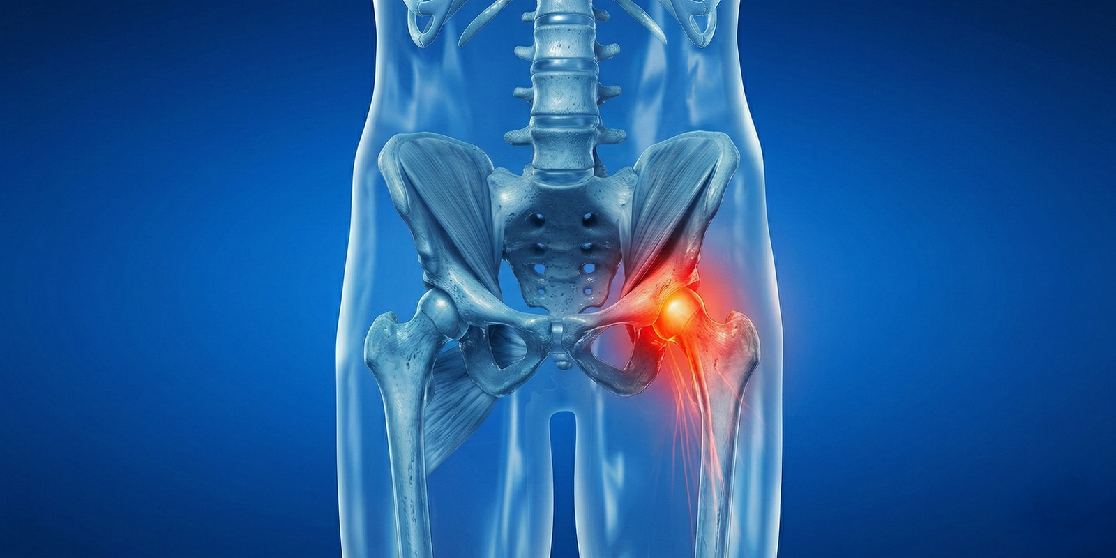
The hip is one of the largest and strongest joints in the body. Walking, running, sitting, standing, and balance all depend on a healthy hip. Its ball-and-socket anatomy allows a wide range of motion and can carry body weight safely. Even so, dislocation can occur under certain conditions. Hip dislocation causes severe pain and is an important orthopedic problem that requires emergency care.
Hip dislocation is defined as complete separation of the femoral head from the acetabulum, the socket in the pelvis. This is not only a change in bone position; the joint capsule, ligaments, muscles, cartilage surfaces, and nearby nerves can also be affected. The sciatic nerve is particularly at risk. If hip dislocation is not treated correctly and promptly, it can lead to permanent loss of function and serious complications.
Why the Hip Does Not Dislocate Easily
The hip stays stable because of bone fit and strong surrounding soft tissues. The femoral head sits deeply in the acetabulum. A thick capsule and ligaments provide high-level stability. The gluteal muscles and deep rotators also contribute.
A healthy hip therefore usually requires high-energy trauma to dislocate. Dislocation is rare in daily activities, but trauma, falls, and accidents can cause fracture and dislocation.
Unlike some other joint dislocations, hip dislocation involves not only bone displacement but also damage to the capsule and surrounding soft tissues. This makes treatment and recovery more complex.
What Is Hip Dislocation?
Hip dislocation is complete separation of the femoral head from the acetabulum. Normally the hip is highly stable because of bone structure, ligaments, capsule, and strong muscles. This stability protects the joint against high loads in daily life. Dislocation therefore usually results from high-energy trauma.
When dislocation occurs, anatomic integrity is lost. As the femoral head leaves the socket, the capsule may tear, ligaments may be injured, and cartilage surfaces may be damaged. Vessels that supply the femoral head may also be stretched or injured. This can later cause impaired circulation and loss of bone vitality. Hip dislocation is therefore considered an orthopedic emergency and should be treated as soon as possible.
What Causes Hip Dislocation?
The most common cause is high-energy trauma. Because the hip is strong, low-energy trauma usually does not cause dislocation. Hip dislocation is therefore most often seen after traffic accidents, falls from height, or serious sports injuries.
Traffic Accidents
Traffic accidents are the most common cause. In vehicle collisions, force transmitted through the knees hitting the dashboard can push the femoral head out of the acetabulum. This is the most common mechanism of posterior hip dislocation. In posterior dislocation the femoral head moves toward the back of the acetabulum.
The patient is usually seen with the leg internally rotated and shortened. This clinical finding is an important diagnostic clue for an experienced physician.
Falls From Height
Construction accidents, falls from stairs, or home accidents from height can overload the hip. In young adults, hip dislocation and associated fractures are often seen after falls from height. Acetabular fractures frequently accompany the dislocation.
Sports Injuries
Sports injuries are less common, but hip dislocation can occur in contact sports or activities that require sudden direction change. Sports-related dislocations are usually linked to high-energy mechanisms and may include fracture fragments in the joint.
Dislocation After Hip Replacement
Dislocation after total hip replacement is increasingly common. In the early postoperative period, muscles may not yet be strong enough and joint stability may not be fully restored, so dislocation risk rises. Not following movement restrictions, crossing the legs, sitting in low chairs, sudden turning, and incorrect lying positions can cause prosthetic hip dislocation.
Recurrent dislocations in a prosthetic hip may require revision surgery depending on component position, soft-tissue balance, and the patient’s general condition.
Structural and Congenital Factors
Structural problems such as congenital hip dysplasia, connective-tissue diseases, and muscle imbalance can reduce hip stability and increase dislocation risk. In these cases the hip is already looser and more unstable, so lower-energy trauma may be enough to cause dislocation.
Types of Hip Dislocation
Hip dislocations are classified by the direction in which the femoral head leaves the acetabulum. This classification is important for treatment planning and complication risk.
Posterior Hip Dislocation
This is the most common type. The femoral head moves toward the back of the socket. Posterior dislocations usually result from traffic accidents and are related to force through the knee. Clinically the leg appears internally rotated and shortened. Sciatic nerve injury risk is higher in this type.
Anterior Hip Dislocation
Anterior dislocations are less common. The femoral head moves forward, usually during excessive opening and external rotation of the hip. Sports injuries and certain trauma mechanisms can cause anterior dislocation. The leg is usually seen in an externally rotated position.
Central (Fracture) Dislocations
In some cases a central dislocation develops together with an acetabular fracture. This is a more complex injury and treatment is more extensive. The femoral head is driven into the acetabulum and the bone may be fragmented.
Prosthetic Hip Dislocations
Prosthetic hip dislocations differ in mechanism from native hip dislocations. They are usually positional and related to movement. Component placement, soft-tissue balance, and adherence to movement restrictions determine risk.
What Are the Symptoms of Hip Dislocation?
Hip dislocation usually presents with sudden, severe pain. The patient feels intense pain in the hip and cannot move the affected leg. Weight-bearing is often impossible. The leg usually rests in an abnormal position.
In posterior dislocations the leg appears internally rotated and shortened; in anterior dislocations it may be externally rotated. Together with the trauma history, this appearance supports the diagnosis.
Limitation of motion is marked, and even the smallest movement increases pain. Swelling, tenderness, and sometimes bruising may be seen around the hip. In some cases nerve injury causes numbness, tingling, or muscle weakness in the leg. If the sciatic nerve is affected, weakness may appear in the ankle and toes.
Hip dislocation can also cause prolonged stiffness and pain because of damage to surrounding muscles and ligaments. Early diagnosis and treatment are therefore critical for both pain control and preservation of function.
Diagnostic Process
Diagnosis starts with history and physical examination. The mechanism of trauma, pain severity, and position of the leg are guiding. Leg-length difference, rotational deformity, and limited motion are assessed.
Imaging is used to confirm the diagnosis. X-ray clearly shows hip dislocation and is the basic method for associated fractures. Computed tomography may be needed in some cases. CT is used for more detailed assessment of cartilage damage and small fractures. CT is also important after reduction to confirm that the hip is anatomically reduced.
In the emergency setting, patients with suspected hip dislocation also need neurovascular assessment. Sciatic nerve function is checked. If sensory loss, reduced motor power, or circulation problems are present, emergency treatment becomes even more critical.
Treatment of Hip Dislocation
Treatment is planned as an emergency. The main aim is to return the femoral head to the acetabulum as soon as possible. This procedure is called reduction. In most cases reduction is done closed, under general anesthesia or sedation.
Muscle relaxation and sedation increase the chance of success. The femoral head is guided slowly and in a controlled way into the acetabulum. After reduction, hip stability is assessed and control imaging is obtained.
If closed reduction fails or fracture fragments are present in the joint, surgery may be needed. The joint is opened, the femoral head is reduced, and necessary repairs are made. Acetabular fractures are stabilized with plates and screws, and cartilage damage is addressed if present.
A similar approach is used for dislocation after hip replacement. Closed reduction is tried first. In recurrent dislocations, component position is assessed and revision surgery is planned if needed. Revision involves repositioning components and restoring soft-tissue balance. Rehabilitation after this type of surgery is longer and more controlled.
Recovery and Rehabilitation After Hip Dislocation
Recovery depends on the type of dislocation, associated injuries, and the treatment used. After reduction, a period of load restriction is usually applied. Crutches or a walker may be needed.
In the first weeks the aims are to control pain, reduce swelling, and preserve range of motion as much as possible. The physical therapy program is arranged according to general condition and dislocation type. Strengthening the muscles around the hip, especially the gluteal muscles and deep rotators, is critical for restoring stability.
In the second stage the program becomes more active. Balance and proprioception exercises, gait training, and muscle strengthening are added. Gait, leg length, and hip rotation are carefully assessed. Rehabilitation may last 6–12 weeks, and in some cases longer, depending on age, general health, and trauma severity.
In patients who dislocate after hip replacement, rehabilitation may be longer. Special exercises to increase soft-tissue stability and adherence to movement restrictions are very important. Because recurrent dislocation risk is present in this group, patient education, correct sitting and standing techniques, adherence to restrictions, and regular follow-up are essential.
Possible Complications After Hip Dislocation
One of the most important complications is avascular necrosis of the femoral head. This occurs when blood supply to the femoral head is disrupted and bone tissue loses vitality. Avascular necrosis may become apparent months after dislocation, but in some cases it appears years later. Long-term follow-up after hip dislocation is therefore necessary.
Cartilage damage, early osteoarthritis, nerve injury, and recurrent dislocation may also occur. If the joint surface is damaged, the risk of post-traumatic arthritis increases. Osteoarthritis can cause pain and stiffness and later increase the need for hip replacement.
Nerve injury, especially sciatic nerve injury, can occur during dislocation. Weakness, numbness, and tingling may appear in the ankle and toes. In some cases nerve injury is temporary; rarely it may be permanent.
After prosthetic hip dislocation, component displacement, loss of soft-tissue balance, and recurrent dislocation are the main concerns. Further surgery may then be needed.
Quality of Life and Long-term Follow-up
Success is not measured only by reducing the dislocation. In the long term, quality of life, pain level, range of motion, and the ability to continue daily activities are the most important indicators. Follow-up is therefore an integral part of treatment.
In patients at risk of avascular necrosis, regular follow-up and imaging are important for early diagnosis. MRI can help assess femoral head blood supply in the early period. Necrosis can then be detected at an early stage and the treatment plan adjusted.
Long-term follow-up also watches for post-traumatic osteoarthritis. If osteoarthritis symptoms increase, quality of life falls and more advanced options such as hip replacement may be considered.
Conclusion
Hip dislocation is a serious orthopedic injury that usually results from high-energy trauma and requires emergency care. Although the hip is one of the strongest and most stable joints, increasing trauma severity can disrupt stability and fully separate the femoral head from the acetabulum. This is not only a mechanical dislocation; it is a multilayered injury that also puts the capsule, ligaments, cartilage, and neurovascular structures at risk.
The most critical factor is time. Reduction in the first hours is decisive for preserving blood supply to the femoral head. Delayed reduction increases avascular necrosis risk and can lead to long-term loss of hip function. Patients with suspected hip dislocation should therefore receive orthopedic evaluation without delay.
Treatment is not limited to reducing the dislocation. Neurovascular status is assessed, associated fractures are identified, an appropriate rehabilitation plan is made, and a long-term follow-up program is arranged. In patients who dislocate after hip replacement, restoring soft-tissue stability and adhering to movement restrictions are especially important. Early diagnosis, correct treatment, and regular follow-up are needed to preserve quality of life. Most patients can return to daily life with appropriate treatment and rehabilitation, but risks are not completely eliminated and regular checks should not be missed.
Frequently Asked Questions

Specialist Opinion
Hip dislocation is a serious orthopedic injury that usually results from high-energy trauma and requires emergency care. Although the hip is one of the strongest and most stable joints, increasing trauma severity can disrupt stability and fully separate the femoral head from the acetabulum. This is not only a mechanical dislocation; it is a multilayered injury that also puts the capsule, ligaments, cartilage, and neurovascular structures at risk.
The most critical factor is time. Reduction in the first hours is decisive for preserving blood supply to the femoral head. Delayed reduction increases avascular necrosis risk and can lead to long-term loss of hip function. Patients with suspected hip dislocation should therefore receive orthopedic evaluation without delay.
Treatment is not limited to reducing the dislocation. Neurovascular status is assessed, associated fractures are identified, an appropriate rehabilitation plan is made, and a long-term follow-up program is arranged. In patients who dislocate after hip replacement, restoring soft-tissue stability and adhering to movement restrictions are especially important. Early diagnosis, correct treatment, and regular follow-up are needed to preserve quality of life. Most patients can return to daily life with appropriate treatment and rehabilitation, but risks are not completely eliminated and regular checks should not be missed.
Contact us to have your symptoms assessed and to learn about treatment options that may be right for you. Get in touch .
Frequently Asked Questions
No. Hip dislocation always requires medical treatment. Waiting for it to correct itself can seriously damage the joint surface and the blood supply of the femoral head.
In most cases closed reduction is enough. Surgery may be needed if there are fracture fragments in the joint, an unstable dislocation, or failed reduction.
This depends on age, the severity of trauma, and the treatment method. Controlled weight-bearing is usually advised for several weeks. Full recovery may take 2–3 months, and in some cases longer.
Yes, especially with a prosthetic hip or structural problems. Recurrent dislocations may require revision surgery.
Low-risk sports may be possible with a physician’s approval. Contact sports and high-energy activities are not recommended. Planning should be individualized after rehabilitation is complete.
Pain duration varies. Pain is severe at first and decreases over time. If cartilage damage or avascular necrosis develops, pain may become chronic.
Yes, it is one of the most serious complications. Early reduction and regular follow-up reduce this risk but do not eliminate it completely.
 WhatsApp
  Facebook
  Twitter
  Linkedin



 +90 505 398 34 34
 doctoryavuzarikan@gmail.com
 Book Appointment / Contact