

What Is Hemangioma (Vascular Birthmark)? Causes, Symptoms, and Treatment



What Is Hemangioma (Vascular Birthmark)?
Hemangioma (vascular birthmark) is a vascular tumor that develops with abnormal proliferation of the cells that form blood vessels and most often follows a benign course. Although it is seen most on the skin, it can also form in muscle, internal organs, and inside bone tissue. Hemangiomas are therefore not only an aesthetic problem, but in some cases a clinical situation that should be assessed from an orthopedic oncology perspective.
Hemangiomas are seen frequently especially in infancy and childhood, but they can also appear in adults. On the skin they are usually noticed as a red, purple, or blue mark or raised area. Deeply seated hemangiomas may not be visible from the outside and often make themselves known with pain, swelling, or loss of function.
Hemangiomas that develop inside bone carry separate clinical importance. These lesions can create appearances similar to bone tumor, bone infarct, or fibrous dysplasia on imaging. Advanced imaging and specialist assessment are therefore essential to prevent the risk of incorrect diagnosis. In the clinical approach emphasized by Prof. Dr. Yavuz Arıkan, it is stated that bone and intramuscular hemangiomas must be approached from an orthopedic oncology perspective.
A large share of hemangiomas are benign and do not turn into cancer. Hemangiomas that grow, cause pain, weaken bone tissue, or put pressure on nerves, however, require treatment. Vascular birthmarks that sit in critical regions such as the spine, hip, and long bones in particular can increase fracture risk and lead to serious loss of function.
In summary, although hemangioma (vascular birthmark) is most often a harmless vascular tumor, when it sits in bone and deep tissues it can create clinical pictures serious enough to be confused with a bone tumor. Early diagnosis and correct specialist assessment are therefore of great importance for safe, effective treatment.
Why Does Hemangioma Form?
Hemangioma (vascular birthmark) is a benign vascular tumor that appears as a result of uncontrolled, abnormal proliferation of the cells that form blood vessels. Although the exact cause of hemangiomas is not fully known today, it is accepted that several biological and genetic factors play a role in the formation process. Clinical observations in orthopedic oncology show that disruption of vascular structure is the basic factor especially in the development of bone and intramuscular hemangiomas.
According to the clinical approach emphasized by Prof. Dr. Yavuz Arıkan, hemangiomas can be much more than a simple skin mark, and the formation mechanism should be assessed at the cellular level.
1. Congenital Vascular Development Disorders
A large share of hemangiomas appear in connection with congenital disorders of vascular structure. Failure of vessels to develop normally in the embryonic period leads to vascular birthmarks that become obvious after birth.
2. Genetic Predisposition
In some individuals the risk of hemangioma developing is higher. In people with a family history, cellular control mechanisms related to vascular structure may be more sensitive.
3. The Effect of Hormones
Estrogen in particular can stimulate proliferation of vascular cells. Hemangiomas can therefore show a greater tendency to grow in women, during pregnancy, and in those receiving hormone treatment.
4. Loss of Control in Vascular Cells
Normally, vascular cells proliferate in a certain order. When this control mechanism is disrupted, vascular cells proliferate in an abnormal way and hemangioma forms.
5. Trauma and Tissue Damage
Some hemangiomas can develop in regions that have previously been struck or injured. Vascular damage after trauma can trigger abnormal vessel growth.
6. Bone and Deep-Tissue Location
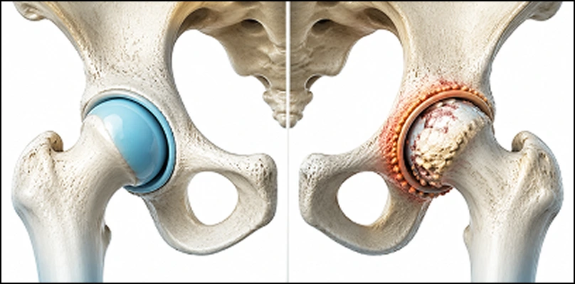
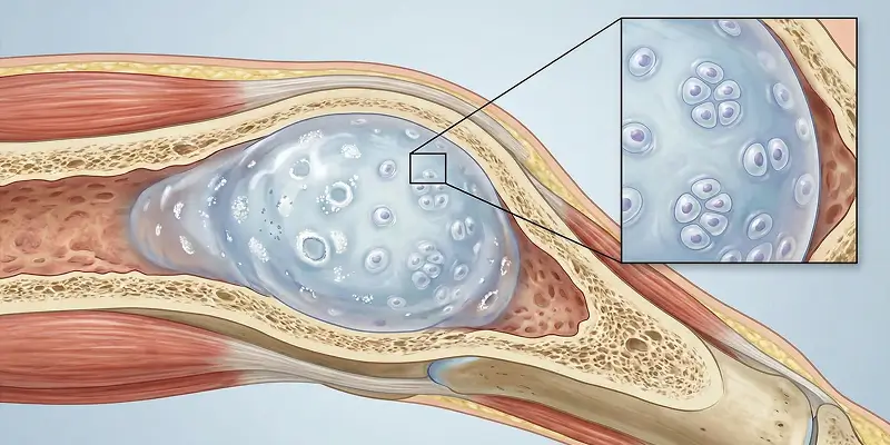
In hemangiomas that develop inside bone, there is abnormal widening of the vascular network inside bone marrow. This situation can create appearances similar to bone tumor and bone infarct and should be assessed carefully from an orthopedic oncology perspective.
In Which Regions Is Hemangioma Seen?
Because hemangioma (vascular birthmark) is a benign vascular tumor originating from blood vessels, it can appear in almost every region of the body that contains vessels. The region where it sits directly affects both the severity of symptoms and the need for treatment. Hemangiomas that sit in deep tissues or inside bone in particular carry great importance from an orthopedic oncology perspective because they can be confused with bone tumor and bone infarct.
According to the clinical experience emphasized by Prof. Dr. Yavuz Arıkan, the location of hemangiomas is one of the most important factors that determine the course of the disease.
1. Skin and Subcutaneous Tissue
The most common place hemangiomas are seen is the skin. They are seen especially on the face, neck, trunk, arms, and legs as a red, purple, or blue vascular birthmark. Superficial hemangiomas are usually noticed early.
2. Muscle Tissue
Hemangiomas that sit inside muscle may not be visible from the outside. These hemangiomas can show themselves with swelling, pain that increases with movement, and muscle weakness, and they can sometimes be confused with a bone tumor.
3. Intraosseous Hemangiomas
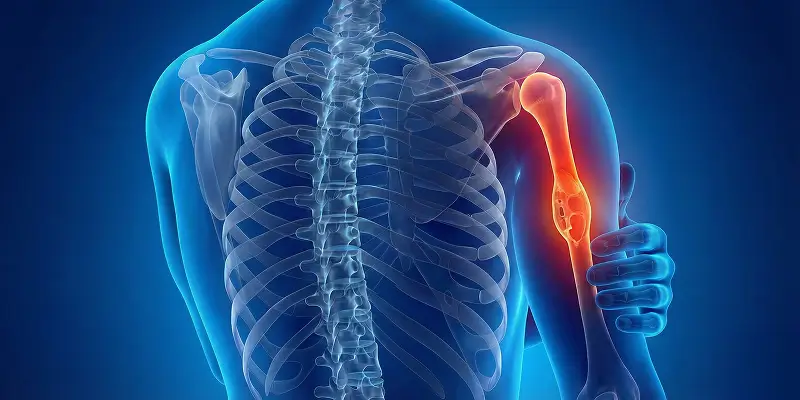
Hemangiomas rarely develop inside bone. The spine, skull, ribs, pelvis, and long bones are affected most. Intraosseous hemangiomas can give appearances similar to bone tumor, fibrous dysplasia, or bone infarct on MRI and CT.
4. Internal Organs
Hemangiomas can also be seen in internal organs. The liver, spleen, and lung are the most frequently involved organs. These hemangiomas are usually found by chance.
5. Near the Spine and Nerves
When spine-located hemangiomas grow, they can cause neurologic symptoms such as back pain, numbness, and loss of strength. Early diagnosis is therefore very important.
What Are the Symptoms of Hemangioma?
Hemangioma (vascular birthmark) symptoms show great differences according to the region where the lesion is found, its size, and its depth. Superficial skin hemangiomas are usually noticed early, while hemangiomas that sit in muscle or inside bone can remain silent for a long time. Bone-located hemangiomas in particular should be assessed carefully from an orthopedic oncology perspective because they can create symptoms similar to bone tumor or bone infarct.
Clinical observations emphasized by Prof. Dr. Yavuz Arıkan show that noticing symptoms in the early period is critical in preventing permanent damage.
1. Color Change on the Skin
The most typical symptom is a red, purple, or blue mark or raised area on the skin. It may fade when pressed and become obvious again when released.
2. Swelling and a Mass
Hemangiomas can be felt as a superficial or deep mass. Deep hemangiomas may not be visible from the outside but give a sense of fullness when touched.
3. Pain
Hemangiomas near muscle, bone, or nerves can cause pain. The pain can be of a type that increases with movement, is felt at night, or is a continuous ache.
4. Increased Warmth and a Pulse Sensation
In some large hemangiomas, because of vascular density, there can be increased warmth in the area and a mild vibration or pulse sensation.
5. Limited Motion
Hemangiomas that sit around a joint or inside muscle can lead to loss of function by preventing movement of muscles and tendons.
6. Findings Related to Nerve Compression
In hemangiomas near the spine or nerves, numbness, tingling, and loss of strength can develop.
7. Bone Weakness and Fracture Risk
Hemangiomas that sit inside bone weaken the bone. This situation can appear with fractures without trauma or sudden severe pain.
The Difference Between Hemangioma and Bone Tumor
Hemangioma (vascular birthmark) and bone tumor are two different clinical pictures that are frequently confused, especially because they can resemble each other on imaging. Their origins, behavior, and treatment approaches, however, are completely different. Making this distinction correctly is of vital importance in orthopedic oncology practice for preventing unnecessary surgeries and planning the right treatment.
In the clinical approach applied by Prof. Dr. Yavuz Arıkan, these two situations are always assessed together with advanced imaging and clinical findings.
1. Difference in Origin
Hemangioma is a vascular, benign formation that develops from the cells that form blood vessels. A bone tumor, on the other hand, forms with abnormal proliferation of cells originating directly from bone tissue or bone marrow.
2. Benign–Malignant Distinction
Hemangiomas are almost always benign and do not metastasize. Bone tumors can be benign or malignant (such as osteosarcoma, chondrosarcoma), and some can threaten life.
3. Imaging Features
On MRI and CT, hemangiomas show a spongy structure with a dense vascular network. Bone tumors are seen as structures that destroy bone, form a hard mass, or disrupt bone tissue. Intraosseous hemangiomas can also sometimes be confused with fibrous dysplasia or bone infarct.
4. Pain and Clinical Course
Hemangiomas most often grow slowly and can remain silent for a long time. Bone tumors usually show themselves with steadily increasing pain, night pain, and swelling.
5. Fracture Risk
Both can weaken the bone, but fracture risk in hemangioma is usually limited. In bone tumors, because bone tissue is destroyed more aggressively, the risk of pathologic fracture is higher.
6. Treatment Approach
Hemangiomas are most often treated with observation or minimal interventions. Bone tumors may require much more aggressive treatments such as surgery, chemotherapy, or radiotherapy. Correct diagnosis therefore prevents treatment from becoming unnecessarily heavy.
How Is Hemangioma Diagnosed?
Hemangioma (vascular birthmark) diagnosis, especially when it sits in deep tissues or inside bone, requires a careful, multi-stage assessment. These lesions can show findings similar to bone tumor, bone infarct, or fibrous dysplasia on imaging. In orthopedic oncology, correct diagnosis is the foundation of preventing unnecessary surgical interventions and planning appropriate treatment.
According to the clinical approach applied by Prof. Dr. Yavuz Arıkan, hemangioma diagnosis is made in a systematic way.
1. Clinical Examination
In the first stage, the lesion’s size, hardness, color change, pulse or increased warmth, and whether it causes pain are assessed. Hemangiomas on the skin are easily noticed visually, while deep hemangiomas can only be felt on examination by hand.
2. Ultrasound
In superficial hemangiomas, ultrasound helps diagnosis by showing vascular structure and blood flow. Vascular structure is shown clearly with color Doppler ultrasound.
3. Magnetic Resonance (MRI)
In deeply seated, intramuscular, or intraosseous hemangiomas, the most valuable method is MRI. With MRI, vascular structure, its relationship with muscle and bone, and distinction from bone tumor, bone infarct, or fibrous dysplasia are made clearly.
4. Computed Tomography (CT)
CT is used especially to see whether bone structure is affected. In intraosseous hemangiomas, widening and thinning in the spongy structure of the bone can be seen.
5. Angiography
In large or complex hemangiomas, angiography can be done to see the source of the vessel and blood flow. This method is important especially in treatment planning.
6. Biopsy
In a large share of hemangiomas, biopsy is not needed. If there is suspicion of a bone tumor on imaging and differential diagnosis is not clear, however, a controlled biopsy is applied by an experienced orthopedic oncology team. Because hemangiomas have a dense vascular structure, this procedure should be performed with special techniques, taking bleeding risk into account.
Treatment Methods for Hemangioma
Hemangioma (vascular birthmark) treatment is determined according to the region where the lesion is found, its size, its depth, and the symptoms it creates in the patient. While many hemangiomas follow a harmless course, some can create pain, limited motion, or fracture risk by showing bone-tumor-like behavior. The treatment decision should therefore be made with an orthopedic oncology approach after detailed assessment.
In the clinical principles applied by Prof. Dr. Yavuz Arıkan, the aim is to avoid unnecessary interventions, but to treat risky hemangiomas in time and in the right way.
1. Follow-Up and Observation
For asymptomatic, small, and superficial hemangiomas, the most frequently preferred approach is regular follow-up. In this process, change in size, change in color or hardness, and development of pain are checked.
2. Medication
In some hemangiomas, drugs that suppress vessel growth can be used. Especially in lesions that show active growth, medication can provide shrinkage.
3. Sclerotherapy (Vessel Closure)
In this method, a special substance is injected into the hemangioma so that the vascular structures close. It is an effective option especially in intramuscular and deep-tissue hemangiomas.
4. Laser and Radiofrequency Treatments
In superficial hemangiomas, laser or radiofrequency methods can be used to shrink or eliminate the vessels.
5. Embolization
In large or densely vascular hemangiomas, the vessels that feed the lesion are closed with special methods. This method is applied especially in spine and intraosseous hemangiomas to reduce bleeding before surgery.
6. Surgical Treatment
Surgery may be needed in the following situations: severe pain, nerve compression, fracture risk, or lesions that cannot be distinguished from a bone tumor. During surgery the hemangioma is completely removed and bone repair is done if needed.
Conclusion
Hemangioma (vascular birthmark), although most often perceived as an innocent skin mark, is in reality a vascular tumor that can sit in the body’s muscle, bone, and internal organs. Especially when it sits inside bone or in deep tissues, it can create appearances that can be confused with serious diseases such as bone tumor, bone infarct, and fibrous dysplasia. Assessing hemangiomas by looking only at their outward appearance is therefore not enough.
In orthopedic oncology practice, in the approach adopted by Prof. Dr. Yavuz Arıkan, the location of the hemangioma, its growth rate, and the effects it creates in the patient are analyzed in detail. Thanks to advanced imaging methods such as MRI, CT, and angiography if needed, the difference between hemangioma and a true bone tumor is shown correctly. Unnecessary biopsies and operations are thus prevented, while risky lesions are also treated in time.
While a large share of hemangiomas can be managed safely with follow-up and observation, in situations that lead to pain, nerve compression, or bone weakening, targeted treatments such as sclerotherapy, embolization, or surgery are applied. This personalized approach both protects quality of life and prevents possible complications.
In conclusion, hemangioma should not be seen as a simple vascular birthmark; especially in bone- and muscle-located cases, it should definitely be assessed from an orthopedic oncology perspective. Thanks to correct diagnosis and appropriate treatment applied at the right time, both the risks hemangioma can create are eliminated and patients can continue their daily lives safely.
Frequently Asked Questions
Frequently Asked Questions
Hemangioma is a vascular tumor that develops with abnormal proliferation of the cells that form blood vessels and most often follows a benign course. It is known among the public as a vascular birthmark.
A large share of hemangiomas are benign and harmless. Hemangiomas that sit in critical regions such as bone or the spine, that grow, or that put pressure on nerves, however, may require treatment.
No. Hemangiomas are almost always benign and do not turn into cancer. They do not metastasize.
Yes. Hemangiomas that develop inside bone can show findings similar to bone tumor, bone infarct, or fibrous dysplasia on imaging. MRI and specialist assessment are therefore required.
Superficial skin hemangiomas are usually painless. Hemangiomas near muscle, bone, or nerves, however, can create pain that increases with movement, night pain, or an aching sensation.
Some infantile hemangiomas can shrink with age. Adult hemangiomas usually do not disappear on their own. They can show a tendency to grow.
It is diagnosed with ultrasound, MRI, CT, and angiography if needed. In deeply seated hemangiomas, MRI is the most valuable method. Biopsy can be done if a bone tumor is suspected.
Small, asymptomatic hemangiomas can be followed. Hemangiomas that grow, cause pain, or weaken bone, however, can lead to fracture risk and loss of function.
These are hemangiomas formed by abnormal widening of the vascular network inside bone marrow. They are seen in regions such as the spine, skull, ribs, and pelvis. They can be confused with a bone tumor.
It is seen most on and under the skin (face, neck, trunk, arms, legs). It can also appear in muscle tissue, inside bone, in the spine, and in internal organs (liver, spleen, lung).
 WhatsApp
  Facebook
  Twitter
  Linkedin



 +90 505 398 34 34
 doctoryavuzarikan@gmail.com
 Book Appointment / Contact