

What Is Chondrosarcoma? Symptoms, Diagnosis, and Treatment



Chondrosarcoma is a type of bone cancer that develops when cartilage cells grow in an uncontrolled, abnormal way in bone and cartilage. These tumors often grow slowly and remain localized at first, but they can later spread into nearby bone, joints, and soft tissue. Chondrosarcoma typically arises in cartilage-containing regions, which distinguishes it from other bone cancers.
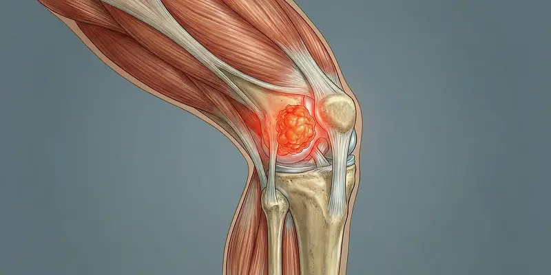
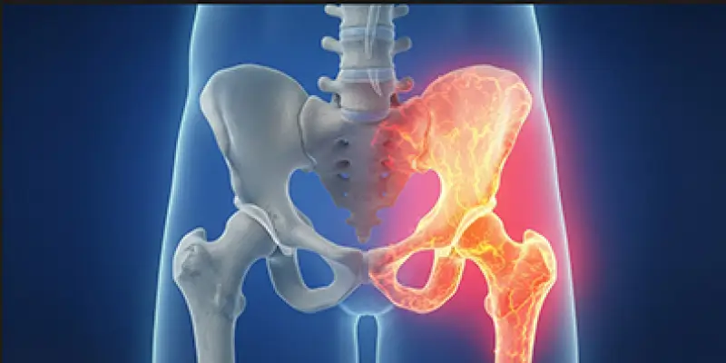
It is most common in the pelvic bones, femur (thigh bone), humerus (upper arm bone), and spine—load-bearing areas rich in cartilage. In these sites it can cause pain, limited motion, and a sense of a mass. Depending on location, it may compress nerves, vessels, or joints and seriously reduce quality of life.
Although uncommon among bone cancers, chondrosarcoma is clinically important because many cases are resistant to chemotherapy and radiotherapy, and surgery is usually the main treatment. Early diagnosis and careful surgical planning are critical for long-term survival.
Most cases occur in people aged 40 and older. It can appear earlier in patients with certain genetic conditions or a history of bone–cartilage tumors. Chondrosarcomas that arise from benign cartilage tumors such as osteochondroma or enchondroma may be recognized late if those lesions are not followed, which can reduce treatment options.
Chondrosarcoma should therefore be managed as a serious oncologic disease that requires accurate diagnosis, experienced surgery, and long-term follow-up—not simply as “a bone tumor.”
Definition of Chondrosarcoma
Chondrosarcoma is a malignant bone tumor that originates from cartilage and can develop inside bone or on the bone surface. Uncontrolled cartilage-cell growth can invade nearby bone, joints, and soft tissue. Because it has features of both bone cancer and a cartilage tumor, it is one of the most important diseases in orthopedic oncology.
Chondrosarcomas are divided into histologic subtypes, and this classification directly affects the course of disease and treatment. The most common and clinically important subtypes include:
Dedifferentiated Chondrosarcoma
This subtype develops when a low-grade chondrosarcoma transforms into a high-grade, aggressive bone cancer. It grows rapidly and has a high risk of local spread and metastasis. Combined surgery, chemotherapy, and radiotherapy may be required.
Myxoid Chondrosarcoma
Cartilage cells form a gelatinous (myxoid) matrix. This type can behave more aggressively than classic chondrosarcoma and may tend to spread into soft tissue.
Secondary Chondrosarcoma
This type arises from a preexisting benign cartilage tumor (such as osteochondroma or enchondroma). Transformation from osteochondroma to chondrosarcoma is a well-recognized scenario in orthopedic oncology practices in Istanbul, and delayed diagnosis is a risk if follow-up is not regular.
Each subtype differs in spread potential, treatment response, and outlook. Accurate biopsy, pathology review, and evaluation by an experienced orthopedic oncology team are essential.
In Turkey, and especially at orthopedic oncology centers in Istanbul, diagnosis and treatment are multidisciplinary. In Prof. Dr. Yavuz Arıkan’s approach, correctly identifying the subtype is a key factor in planning surgical margins and additional therapy.
Chondrosarcoma Symptoms
Symptoms usually appear slowly and insidiously. Patients often ignore complaints for a long time, and diagnosis is delayed. Because chondrosarcoma is a bone cancer, catching it early directly affects surgical success and survival. Tumors in the pelvis, spine, femur, and humerus may begin with mild symptoms.
The most common reasons patients seek care are:
Pain
Pain is the earliest and most important symptom. It may start mild and intermittent, then become constant and worse at night. It usually does not resolve with rest and, unlike ordinary muscle–joint pain, steadily intensifies. In orthopedic oncology clinics in Istanbul, night-worsening bone pain is considered an alarm finding for chondrosarcoma.
Swelling and a Sense of a Mass
As the tumor grows, a visible swelling or firm mass may appear on the bone surface or in nearby soft tissue. The swelling is often painful and tender to pressure. This is more noticeable in cartilage-rich regions.
Limited Motion
If the tumor is near a joint, movement becomes increasingly difficult. Patients may struggle to raise an arm, walk, or sit and stand. Chondrosarcomas around the knee, hip, or shoulder can cause significant loss of function.
Persistent Discomfort
Some patients describe pressure, fullness, or unease inside the bone rather than clear pain. This feeling lasts a long time and may later become pain. It is common in cartilage-origin bone tumors.
Advanced-Stage Symptoms
As chondrosarcoma progresses, additional signs may include:
- Increased warmth in the affected area
- Bone weakness and pathologic fracture
- Numbness or weakness from nerve compression
- In the spine, marked back pain or paralysis findings
These signs may indicate spread into surrounding tissues and a more aggressive stage.
As Prof. Dr. Yavuz Arıkan emphasizes, night-worsening bone pain, a growing swelling, and limited motion together should raise concern for chondrosarcoma or another bone cancer—not a simple orthopedic problem.
Diagnostic Methods
Diagnosis is not based on a single scan. Clinical findings, imaging, and pathologic confirmation are assessed together. Chondrosarcoma can resemble benign cartilage tumors (osteochondroma, enchondroma), but treatment is entirely different. At orthopedic oncology centers in Istanbul, diagnosis is therefore multidisciplinary.
Key methods include:
X-ray (Plain Radiograph)
X-ray is the first imaging study when chondrosarcoma is suspected. Findings may include:
- Calcifications typical of cartilage-origin tumors
- Lytic (bone-destroying) areas
- Cortical thinning or destruction
Borders are often irregular, which helps distinguish chondrosarcoma from benign cartilage tumors.
Magnetic Resonance Imaging (MRI)
MRI is the most critical imaging study. It clearly shows:
- True tumor size
- Spread within the bone marrow
- Proximity to muscle, vessels, and nerves
This information is vital for surgical planning. In Prof. Dr. Yavuz Arıkan’s approach, MRI findings determine the margins of resection.
Computed Tomography (CT)
CT is used for detailed bone assessment. It shows:
- Cortical destruction
- Whether the tumor has extended outside bone
- Calcification patterns
Ring-like calcifications are typical of chondrosarcoma.
PET-CT and Bone Scintigraphy
In some cases, PET-CT or bone scintigraphy is used to look for metastasis. These studies are especially important in high-grade chondrosarcoma.
Biopsy (Definitive Diagnosis)
Imaging may suggest chondrosarcoma, but biopsy confirms it. Pathology determines:
- Whether the tumor is truly chondrosarcoma
- The subtype
- The grade (low, intermediate, or high)
This information decides whether surgery alone or additional treatments are needed.
Incorrect or poorly controlled biopsies can spread tumor cells, so the procedure must be done at centers experienced in sarcoma and bone tumors.
Treatment
Treatment is planned according to tumor size, location, histologic grade, and the patient’s overall health. Unlike many bone cancers, chondrosarcoma of cartilage origin responds only modestly to chemotherapy and radiotherapy. Surgery is therefore the foundation of care in most cases. Radiotherapy and chemotherapy are used as support in some aggressive or advanced tumors.
Main methods include:
Surgery
Surgery is the most effective and critical treatment. The tumor is removed with the widest safe margins possible. Chondrosarcoma cells may have spread microscopically into nearby bone, and inadequate margins can lead to recurrence.
The goals of surgery are:
- Complete clearance of tumor tissue
- Preservation of limb function
Depending on size and location, reconstruction may use:
- Bone grafts
- Specialized tumor prostheses
- Endoprosthetic reconstruction
Many patients can continue life without losing an arm or leg.
In orthopedic oncology teams in Istanbul, Prof. Dr. Yavuz Arıkan’s aim is not only to remove the cancer but also to preserve walking, grasping, and daily function as much as possible.
Radiotherapy
Radiotherapy is usually given after surgery as an adjunct. Although chondrosarcoma cells are relatively radioresistant, radiotherapy is useful when:
- The tumor cannot be completely removed
- Surgical margins are at risk
- The tumor is in a critical site such as the spine or skull base
- Surgery is not possible in advanced disease
The aim is to suppress residual cancer cells, reduce recurrence risk, and slow tumor growth.
Chemotherapy
Chemotherapy is usually not first-line treatment for classic chondrosarcoma, because the cells are largely chemoresistant. It may be considered for:
- Dedifferentiated chondrosarcoma
- High-grade aggressive subtypes
- Spread to the lungs or other organs (metastasis)
In these cases, chemotherapy is used to reduce tumor burden, control spread, and support surgery.
Prognosis (Survival and Chance of Recovery)
Prognosis depends on histologic type, grade (low versus high aggressiveness), size, stage at diagnosis, and the success of treatment. Chondrosarcoma does not progress the same way in every patient; some types grow slowly for years, while others spread rapidly.
In general:
- Low-grade chondrosarcomas grow slowly, have a low chance of metastasis, and, when completely removed with appropriate surgery, allow many years of healthy life.
- High-grade chondrosarcomas are more aggressive and may spread to nearby tissues and especially the lungs. In these cases, both survival and recurrence risk are less favorable.
Main factors affecting prognosis:
- Tumor size
- Location in difficult sites such as the pelvis or spine
- Whether the tumor was removed with clean margins
- Presence of metastasis
- Dedifferentiated or high-grade histology
Survival rates are quite high in patients with low-grade chondrosarcoma who are diagnosed early and treated with appropriate surgery.
Overall Course of the Disease
Chondrosarcoma is a rare bone and cartilage tumor, but if it is not managed correctly it can cause serious loss of function and risk to life. Because symptoms start slowly, many patients already have a sizable tumor at diagnosis, which can make surgery more difficult.
However, when:
- Diagnosis is early
- An experienced orthopedic oncology team is involved
- Surgical margins are appropriate
most patients can continue life without limb loss and with long-term disease control.
As Prof. Dr. Yavuz Arıkan emphasizes, success in chondrosarcoma is not only about the day of surgery; it depends on years of follow-up. Regular imaging and clinical visits are essential for catching recurrence early.
Frequently Asked Questions
Frequently Asked Questions
Not every chondrosarcoma is fatal. Low-grade tumors can often be fully controlled with early diagnosis and appropriate surgery. High-grade and metastatic types carry a higher risk to life.
Yes. Benign cartilage tumors such as osteochondroma and enchondroma can transform into chondrosarcoma over time. These lesions must be followed regularly.
Yes. Recurrence is more likely if surgical margins are inadequate or the tumor is aggressive. Long-term MRI, CT, and chest imaging after surgery are essential.
Chondrosarcoma cells are more resistant to chemotherapy than many other bone cancers. Chemotherapy is used mainly in dedifferentiated or metastatic cases.
With modern techniques, the tumor can often be removed while preserving the limb. Endoprostheses and bone reconstruction are used for this purpose.
It most often metastasizes to the lungs. Less commonly, it can spread to the liver and other bones.
Yes. Especially low-grade chondrosarcomas, when completely removed with appropriate surgery, can allow patients to live disease-free for many years.
 WhatsApp
  Facebook
  Twitter
  Linkedin




 +90 505 398 34 34
 doctoryavuzarikan@gmail.com
 Book Appointment / Contact